How animals sense Earth’s magnetic field is one of biology’s enduring mysteries.
Researchers in Science have now identified superparamagnetic macrophages in the livers of rock pigeons to be crucial for magnetic sensing. The finding uncovers an unexpected role for immune cells in sensory perception and may fundamentally change our understanding of animal navigation.
Learn more in this week's issue: https://t.co/JS9qBFZHcP
Why are there intronic reads in your bulk RNA-seq data?
You're not alone—it's common, and the reasons are more layered than you think.
Let’s break it down. 🧵
🎉 Excited to share that the Danioheart Atlas has now been published! 🎉
We’ve mapped zebrafish heart development across ten developmental stages.
📖 Read the full paper here: https://t.co/6BeSPwCzXE
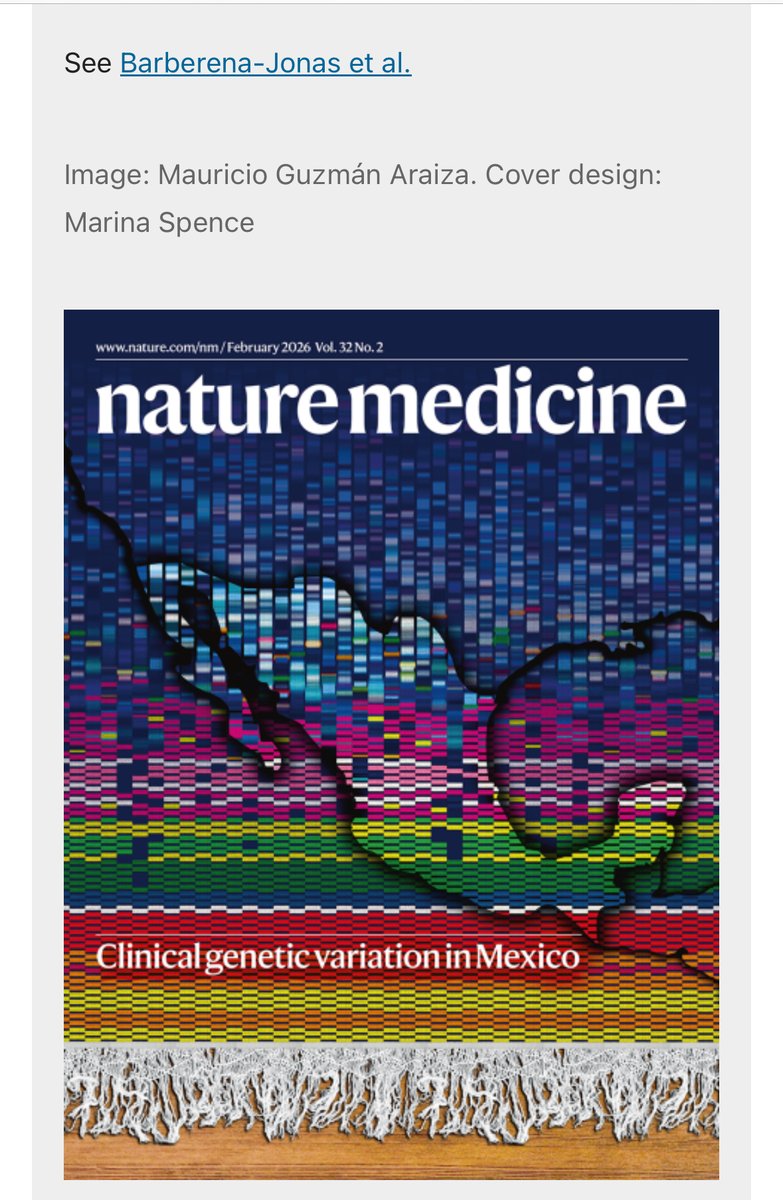
Second time the MX biobank project (https://t.co/qaLnfPzcz2) gets the cover in a @Nature journal! This time in @NatureMedicine highlighting the work by @BarjonCar on Clinical genetic variation in Mexico. Another amazing cover by @mauguz33! https://t.co/k3eGLX4809
How to locate a drug through the entire body.
A new technique, vCATCH, was used to visualize where cancer drugs bind throughout an entire mouse at single-cell resolution. This seems like a useful way to figure out why some drugs cause unintended side effects, or to refine drugs to target tissues more precisely.
There are other ways to track where cells go in the body, of course. For example, drugs can be tagged with radioactive labels and then radioactive imaging used to quantify where the molecules accumulate. But this is not single-cell resolution.
For this paper, the authors studied two cancer drugs: Afatinib, which targets EGFR and is used to treat non-small cell lung cancer, and Ibrutinib, which targets BTK and is used to treat some blood cancers. Both of these drugs can cause brutal side effects (like skin reactions, jaundice, and other fun things) and both are covalent drugs, meaning they form permanent, atomic bonds with their protein targets.
The vCATCH method is really clever. It makes use of click chemistry, a type of reaction used to fuse molecules together using a copper catalyst. Without this copper catalyst, the reaction happens extremely slowly. At a high level, vCATCH works like this:
> Take a drug and chemically modify it to carry an alkyne group (a carbon-carbon triple bond.) This doesn’t usually change how the drug behaves in the body, but it could in some cases.
> Give the modified drug to mice (or another animal.) Wait. The drug binds to its target.
> Kill the animal. Keep it intact.
> “Clear” the tissue. In other words, literally make it more transparent. This is done using a method called HYBRiD clearing. There are several steps here, but the gist is that you want to eliminate lipids because they scatter light. A solvent, usually tetrahydrofuran, is used to do this.
> Drench the cleared tissue with copper. Let it seep into the tissue. (Remember that copper is needed for the click reactions to happen.)
> Add fluorescent dye molecules with azide groups (nitrogens that react with alkynes) to the tissue. These molecules seep inside and “click” together with the drug’s alkyne group.
> The tissue now has a bright, fluorescent dot everywhere the drug is located.
> Use lightsheet microscopy to locate the drugs’ locations.
The beauty of this method is that you can quantify not only how much of the drug went to each organ, but also which cell types in each organ are actually being targeted.
So with Afatinib, for example, they found that it went all over lung tissue (where EGFR is highly expressed; no surprise here), but also to glomeruli in the kidney’s cortex and to particular blood vessels in bone and the spleen. These targets were not previously known, and they might explain some of this drug's side effects or help other biotechs develop more precise variants.
This is a really nice paper. Recommend.
This is a rare find - a beautiful sapphirinid copepod from the waters of the Lombok Strait, Indonesia. This whole animal is only 3 mm in length, fully transparent. They are known for their striking, iridescent colors, which are only displayed by the males.
La Secretaría de Relaciones Exteriores informa que, hasta el momento, se ha confirmado que las embarcaciones en las que viajan Arlin Medrano Guzmán, Sol González Eguía y Carlos Pérez Osorio de nacionalidad mexicana, como parte de la Flotilla Global Sumud, fueron interceptadas por autoridades de Israel y se encuentran en traslado al puerto de Ashdod.
Los servicios consulares de la Embajada de México en Israel estarán presentes y atentos a su llegada para ofrecer la protección y asistencia que requieran. El embajador de México en Israel, Mauricio Escanero, ha estado personalmente en comunicación, hasta el día de hoy, con las personas mexicanas que viajan en la flotilla.
La SRE reitera su llamado a respetar su integridad física y su seguridad, que puedan acceder de manera expedita a la entrevista consular y proceder a su rápida repatriación, en apego al derecho internacional, en particular el derecho internacional de los derechos humanos y el derecho internacional humanitario.
A single aquaporin protein lets one billion water molecules into a cell each second. It is one of the "fastest" channels in all of biology.
Each water flows through in single file, while H+, sodium, potassium, and other ions are excluded.
What explains this remarkable selectivity? Two clever protein filters.
But first: Each aquaporin protein is made from six alpha-helices that run back and forth through the cell membrane. These helices form an hourglass shape so that the channel is cone-shaped at either end and extremely narrow in the middle.
The two ends of the protein also mirror each other, with their point of symmetry occurring at the narrowest part of the channel. It's thought that modern aquaporins evolved through a gene duplication event.
Now let's zoom in a bit and look at the two filters within aquaporin that account for its extreme selectivity. Water can flow either way through the channel, so we'll imagine that we're moving from OUTSIDE the cell to INSIDE the cell.
The first filter we encounter is called the ar/R filter. This is a cluster of polar and charged amino acids that point inside the aquaporin channel.
These four amino acids are *usually* arginine (+ charge), histidine (polar), and two bulky amino acids like phenylalanine and tryptophan.
These four amino acids come together to restrict the middle of the pore, making it just wide enough for a single water molecule to pass. The positively charged arginine also helps "stabilize" water molecules while forcing away sodium ions or protons.
The second filter encountered on our journey into the cell is called the NPA motif (short for asparagine–proline–alanine). There are two of them inside each aquaporin.
These two protein motifs point at each other in the center of the aquaporin. As a water molecule passes the ar/R filter and moves deeper into the channel, it forms hydrogen bonds with the asparagine amino acid in the first NPA motif.
But because the two motifs are oriented in opposite directions, the local electric field at the channel’s midpoint next forces each water molecule to rotate 180° and latch onto the asparagine in the second NPA motif. In other words, each water molecule gets flipped around inside the aquaporin channel.
This "flipping" is important because it breaks the water molecules free from any hitch-hiking protons. It acts as the final filter.
In the early 1940s, there was intense debate about the “true nature” of bacteriophages, the little viruses that infect bacteria.
Some biologists argued that they were bacterial enzymes, whereas others believed they were their own viral entities.
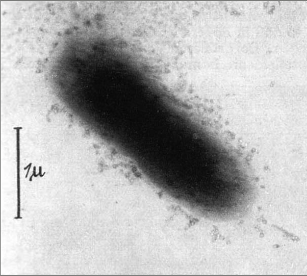
In 1940, Ernst Ruska (the same person who invented the electron microscope in 1931), published an article in German showing the first images of a bacteriophage.
Ruska’s original electron microscope magnified objects only about 400x, much less than light microscopes available at that time. But by 1940, advances by University of Toronto scientists pushed that magnification up to 7,000x, or about 3x higher than light microscopes were then capable of.
Using one of these newer electron microscopes, Ruska captured his photos of bacteriophages. Unfortunately, the images were not so great. It was difficult to tell, for example, whether these were well-defined particles or just random debris from the bacterial cell. (Ruska’s image is the first one below.)
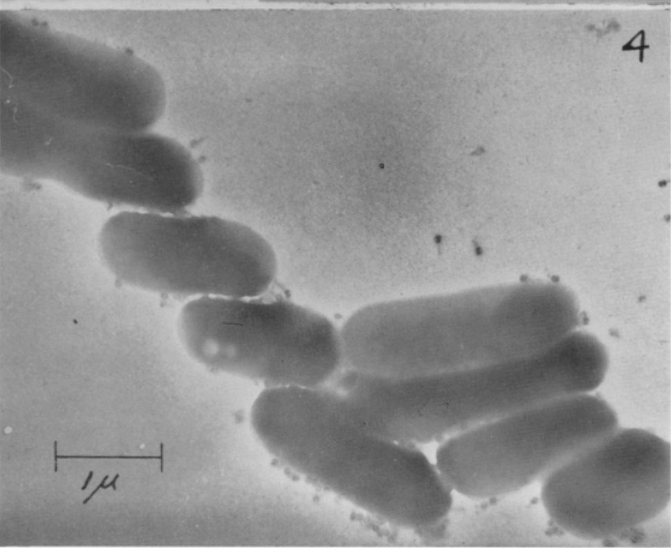
In 1942, two Americans — the brilliant Salvador Luria and Thomas Anderson, both at Columbia University — finally settled the debate.
Using a RCA Electron Microscope (of which only 2,000 units were ever made, each costing about $200,000) the two men acquired much better images of bacteriophages nestled upon a single E. Coli cell. (Their image is the second one below).
With this image, they could clearly see individual phages and their little tails. They saw, too, that these phages were of an “extremely constant and characteristic aspect,” meaning they could not just be random cell debris or enzymes (since the phages had two parts; heads and tails).
Their experiment worked like this: The duo dropped some phages on a tiny collodion film (made of cellulose, and thin enough not to distort the electron beams too much), put them into the machine, and then used a vacuum pump to suck air out of the column. (Without the vacuum, electrons would bounce off air particles and scatter.)
Next, they aligned the focus using a fluorescent screen on the front console. This fluorescent plate would convert the invisible electron image into visible light so the operator could see and tune the image live.
Finally, taking a micrograph involved opening a shutter for a second or two, then closing it and resetting the system for another shot. There was a long glass plate, at the bottom of the column, that caught the electrons which scattered off the phages. Each glass plate carried multiple small frames, so a session could produce several images before the plate had to be removed and developed in a darkroom.
It’s wild to me that these images were taken in the early 1940s; or that engineers were able to build these half-ton, ten-foot-tall machines that could blast biological samples and resolve their structures at such high resolutions. Brilliant.
Del hecho ocurrido esta tarde en Iztapalapa, informo que, al corte de las 22:30 horas, lamentablemente suman cuatro las personas fallecidas; registramos 90 lesionados y, gracias al trabajo de las y los médicos, se han dado de alta a 10 personas.
A través de la CEAVI desplegamos equipos en todos los hospitales para contactar a las familias de las víctimas y brindar el apoyo correspondiente.
Continuaremos con la atención por este lamentable hecho y seguiremos informando.
Para información de personas lesionadas ponemos a disposición el número 55 5683 2222
SCIENCE IN MEXICO: A RISING FORCE AMID ADVERSITY. I came back knowing the funding would be scarce, the bureaucracy maddening, and the equipment overpriced. But also knowing I’d find freedom, brilliant students, and generous colleagues. I have no regrets https://t.co/axIr4UJvo3
¿Por qué pensamos a los espacios verdes como sitios repletos de pasto, sin "malezas"? ¿Para quiénes son realmente estos espacios? ¿Privilegiamos al ornato y "orden" sobre lo social y ecológico?
De esto escribo en mi columna para @RevistaEstePais 🌳🌱
https://t.co/I71EUpv2B3
How do cells clean up misfolded proteins when they divide?
This study finds that the cleanup crew includes the ER chaperone BiP and the proteasome, and kicks in right as cells finish dividing. Surprisingly, one major cell cycle regulator isn’t involved.
https://t.co/cabMJXGUNO