La prohibición de fumar, a la gente le parece bien porque es un tema que se refiere a la salud. Es lo que se llama «estado terapéutico» o «farmacracia»: la idea de que, en vez de justificarse en la defensa, se justifica en la salud. (1/8)
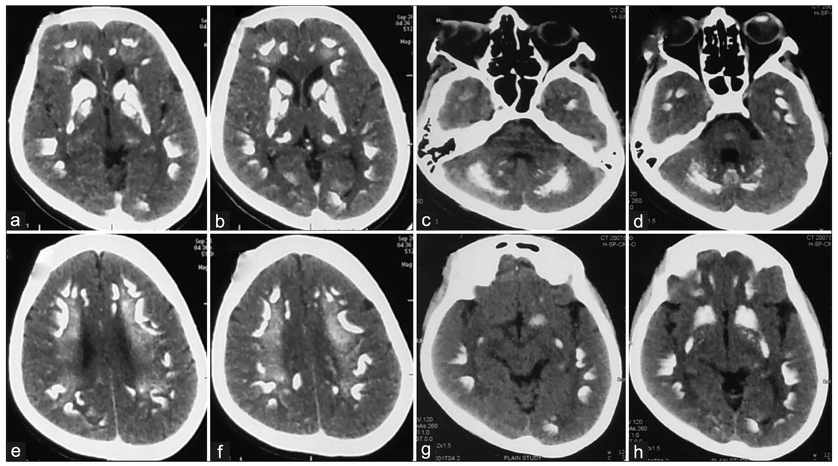
Isolated hemichorea in adults can be a presenting feature of hypoparathyroidism with B/L basal ganglia calcification. Treatable with correction of underlying metabolic abnormality. Movement disorder + brain calcification -> r/o hypoparathyroidism
Link: 10.4103/aian.AIAN_123_18
La representación nacional no es un concepto que me acabe de convencer. Es como la transustanciación: coges a cualquiera, lo unges con votos, y se convierte en representante de la nación. ¿Estamos de broma o qué? (1/6)
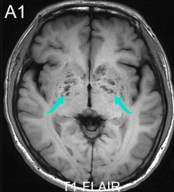
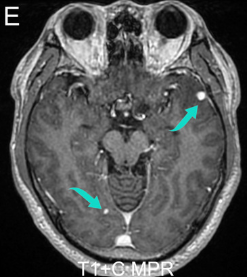
🩺Clinical & neuroimaging 🧠 clues to differentiate 🧩autoimmune encephalitis & its mimics🔎
Current opinions in Neurology 📖
10.1097/WCO.0000000000001489.
No hay ninguna manera sencilla o eficiente de hacer esta devolución del IVA a los estratos populares.
Esto terminará en un berenjenal donde prevalecerá la corrupción y los pobres siempre pagando el aumento del IVA a la canasta básica.
Pondering how to remember pontine syndromes?
Don’t become despondent about syndromes of the pons!
Here’s an easy way remember the basic parts of pontine syndromes:
Pontine lesions are usually vascular and are either from an infarct in the AICA territory (lateral) or paramedian branches of the basilar artery (medial)
Lateral & medial pontine syndromes can be remembered by the easy mnemonic THE PONS, w/ “THE” representing lateral syndromes & “PONS” representing medial syndromes
LATERAL SYNDROMES:
T = Temperature (spinothalamic = loss of pain & temp contralateral body, trigeminal nucleus = ipsilat face)
H = Horner’s (sympathetic pathway)
E = Equilibrium problems (spinocerebellar = ataxia of ipsilateral arm & leg)
MEDIAL SYNDROMES:
P = Proprioception (medial lemniscus = loss of contralateral vibration & proprioception)
O = Ophthalmoplegia (MLF = internuclear ophthalmoplegia)
N = Nonmobile (corticospinal tract = weakness of contralateral body)
S = Sixth nerve (Sixth nerve nucleus = loss of ipsilateral CN 6)
There are many different syndromes that vary by what exact structures are involved, but if you always remember THE PONS, you can easily pontificate about pontine lesions!
"Asymmetric Petro-Occipital Venous Plexus Thickening: A Valuable Diagnostic Sign of Reduced Flow in the ICA"
https://t.co/wLOkxh1V0X
@WassermanBruce@JennyYang189132
This chart summarizes the Oxfordshire Community Stroke Project (OCSP) classification system, also known as the Bamford Classification.
This system categorizes ischemic strokes into four clinical syndromes based entirely on initial presenting symptoms and signs, correlating directly with underlying vascular anatomy and characteristic CT scan features.
https://t.co/2Kfir0wl3W
12/So now you know how to remember all the lobules of the vermis and have a mnemonic to remember their order.
May your knowledge continue to culminate and never declive!
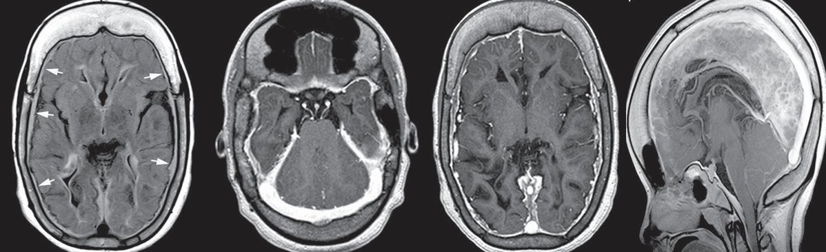
🤵A 37-year-old man presents with 3 weeks of progressive headache, intermittent fever, vomiting, and recent confusion. Brain MRI is performed after clinical deterioration.
🏥What is the most likely diagnosis?
Most people (including plenty of radiologists) misunderstand "resolution" in medical imaging, and the recent @midjourney debate hasn't helped. What resolution actually is, why your scanner's limit is set on day one, and the limit set by a Swedish-American named Nyquist 🧵1/10.
RCVS causes headache, seizures, focal deficits & encephalopathy. Sylaja et al presented the largest single series of RCVS from India (n=22), with favorable short‑term outcome. Most common vascular abnormality was diffuse vasospasm, but focal in 25% Link: 10.4103/aian.aian_667_24
"Atypical Imaging Manifestations of Lateral Dural Tears with Arachnoid Herniations: A Multi-Institutional Study"
https://t.co/CAVanUxPJP
@AjayMadhavanMD
This week's SPIN-POV: Along with slit ventricles and subdural collections, chronic CSF overdrainage after shunt surgery can produce imaging features of intracranial hypotension. Recognizing this constellation is important, as severe cases may require shunt revision.
Having trouble remembering what to look for on scans for Alzheimer’s dementia?
Want the key findings to stay in your memory?
Medial temporal atrophy (MTA) score quantifies hippocampal volume loss
It rates the amount of CSF around the hippocampus as a marker of volume loss
CSF surrounds the hippocampus like a snake surrounding its prey—how far the snake has gone in getting its prey determines the MTA score:
MTA-1: Choroidal fissure opens, looks like the snake has started to slither in
Remember: the number one looks flat like a slightly opened choroidal fissure
MTA-2: Temporal horn widens, looks like the snake has surround the hippocampus
Remember: the number two look like the CSF curving around as the temporal horn opens
MTA-3: Moderate hippocampal atrophy, looks like the snake has begun to squeeze the hippocampus
Remember: the number three has a tight curve, so it looks like it is crushing the hippocampus & it looks small (atrophied)
MTA-4: Severe hippocampal atrophy, looks like the snake has crushed the hippocampus
Remember: Number four has only a tiny hole in it, so the hippocampus has to be super tiny & atrophied to fit inside!
Hopefully, this will help you commit this rating system to memory so you’ll never forget what you need to say for Alzheimer’s!
💉🧠Study finds IVT+EVT for medium distal vessel occlusions was associated with worse functional outcomes vs IVT alone
👉Supports recent research but more RCTs needed
Read more here👇
https://t.co/R5wtmHeSck