🎉This month @naturecvr celebrates the female scientists that published with us last year, by reporting a brief story of their scientific and life journey. Here, we present to you Luisa Iruela Arispe @mliarispe#internationalwomensday#CardioTwitter
👉https://t.co/dtuwYnkpfm
Ruixuan Gao, AP of @ChemistryatUIC and @UIC_BioS at #UICLAS, is one of three researchers recently selected to receive funding from the McKnight Endowment Fund for Neuroscience! Congratulations, Professor Gao!
Read more: https://t.co/6e7rODZGFg
Nikon Imaging Center at the Feinberg School of Medicine in Chicago is looking for a manager. Please look at the announcement at https://t.co/raertcvBTm and email your application to [email protected]
Please RT
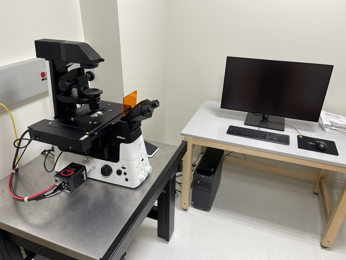
It took a while (pandemic, war, chip crisis, etc.) but finally it is here! Check out our new TIRF microscope - Nikon eclipse Ti2-E equipped with a Photometrics BSI camera. Let the fun begin!🔬🤓😍
@biumedicine@Bar_ilan@NikonInst#microtubules
Research from Prof. Roy Soberman and colleagues uses #SuperResolution#Microscopy with a @Yokogawa_LS SoRa system to help show how liquid-liquid phase separation induced by transcription inhibition results in active chromatin being localized to nucleoli: https://t.co/vpcOy2wHlr
It's such a great honor to be part of the 2022 Searle Scholars cohort! I'm extremely grateful to my mentors, collaborators, colleagues, staff, and lab members, who continue to support our science and passion. @ChemistryatUIC@UIC_BioS@thisisUIC
(1/2) First preprint from the lab! We found that PFA fixation can both enhance and diminish LLPS in cells, even causing puncta to artificially appear or disappear. Our work suggests caution in claiming in vivo LLPS based solely on fixed-cell imaging like immunofluorescence.
Our recent #Microscopy#AppNote details a #HighContent long-term apoptosis assay using phase #Imaging with the Nikon Volume Contrast system to identify single cells, with classification as apoptotic or dead based on readout from fluorescent sensors:
https://t.co/sJiwyuPKH4
Whole slide of spinal cords imaged today at the @NikonInst AXR demo. It’s been a great demo with stellar samples from some excited labs at @JeffersonUniv. Video shows 2x6 fields imaged through the new PA lambdaD 2x with the updated 2k resonant scanner.
Congratulations to members of the Goldman Lab @IFilaments for new research in PNAS, “Nuclear lamin isoforms differentially contribute to LINC complex-dependent nucleocytoskeletal coupling and whole-cell mechanics.” @vahabikashi@SuganSivaguru @FSMsadam
https://t.co/7O37DZu6zS
Check out this @bioprotocolbyte detailing the creation of 3D collagen I/fibronectin matrices for #Imaging cancer cell migration – demonstrated in part using a Nikon A1R #Confocal#Microscopy system: https://t.co/FoCdEBbrV7
Click and check out Nikon #Microscopy product solutions for the cell/tissue culture laboratory, including our ECLIPSE Ts2 line of inverted tissue culture microscopes: https://t.co/eYK91Rkwo7