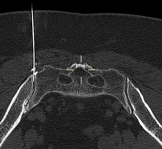
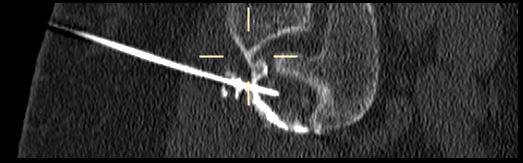
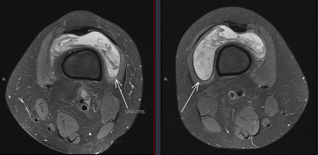
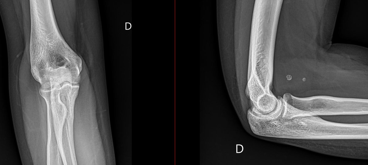
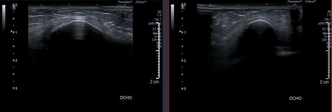
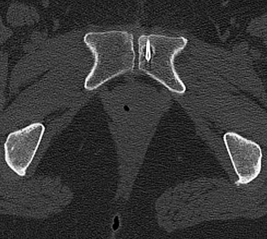
20/9/2023. We performed a CT-guided right sacroiliac joint therapeutic-diagnostic infiltration with steroids and anesthetics, adding iodine contrast to confirm intraarticular treatment. Prone position. #MSK#rehabilitation#traumatology#pain#CT#intervention
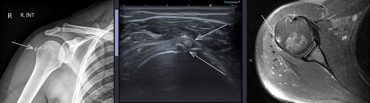
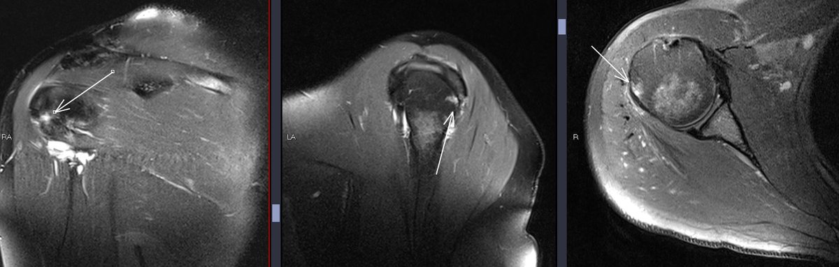
11/9/2023. Evolution of calcifying tendinopathy of teres minor from type I (X-ray) to type II (US) and type III (MRI), with subtle bone marrow edema. 36 yo female painful shoulder. #shoulder#tendinopathy#msk#ultrasound#xray#MRI
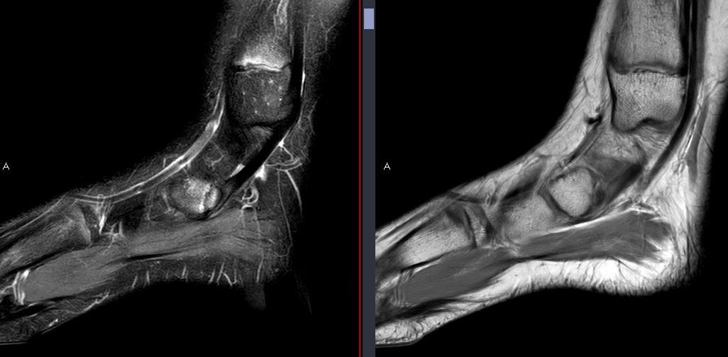
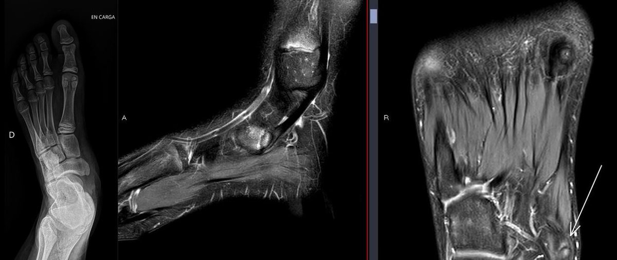
8/9/2023. 13-year-old with medial midfoot pain. Signs of traction apophysitis of the scaphoid's medial ossification center, pulled by the posterior tibialis tendon, with bone marrow edema on both sides of the synchondrosis. 🦶🧒💡 #PediatricOrtho#MSKRadiology#Apophysitis
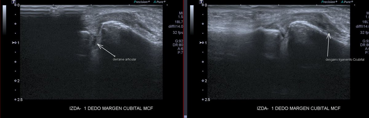
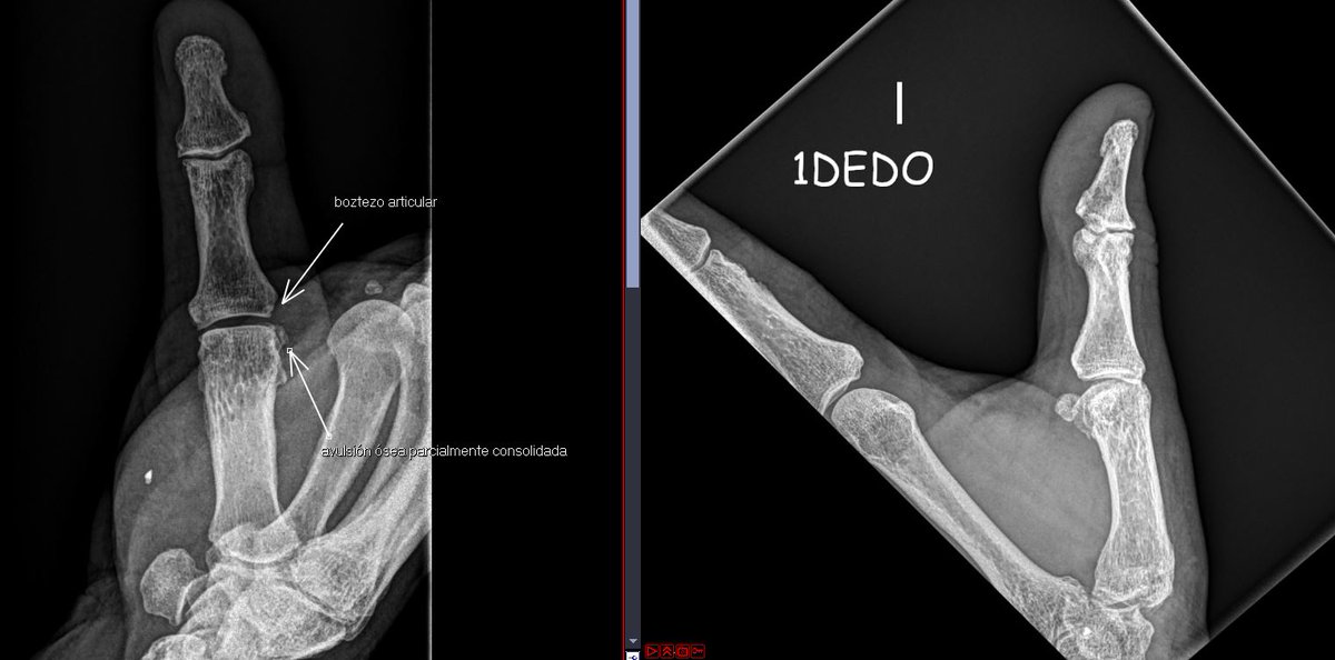
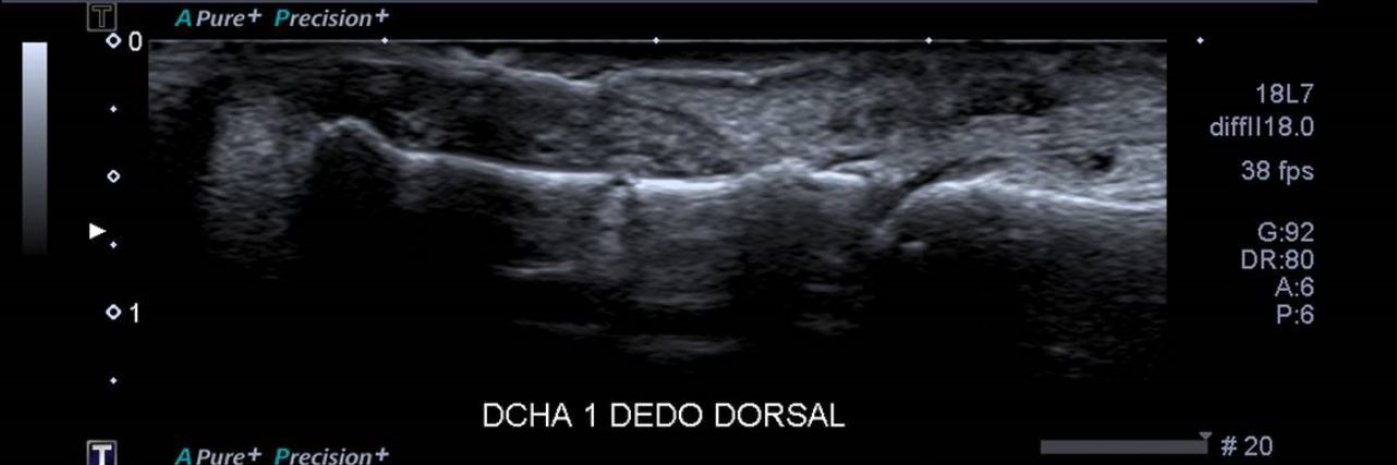
8/9/2023. Mountain bike accident = Mild instability and pain during forced abduction of the thumb. Diagnosis: Proximal bony avulsion of the ulnar collateral ligament of the first MCF joint + no interposition of the adductor pollicis aponeurosis -not a Stenner lesion-#ultrasound
Consent ✅
Runner
Pain ++ 3rd intermetatarsal space, radiating into 2nd and 3rd toes
US - large bursitis & neuroma nestled between 2nd & 3rd met heads (see longitudinal section panning through side-to-side)
US guided CSI - in plane longitudinal approach
Can see the bursal sac filling beautifully - 'whirlpool' effect (taking care to avoid vessel seen pulsing L side of screen)
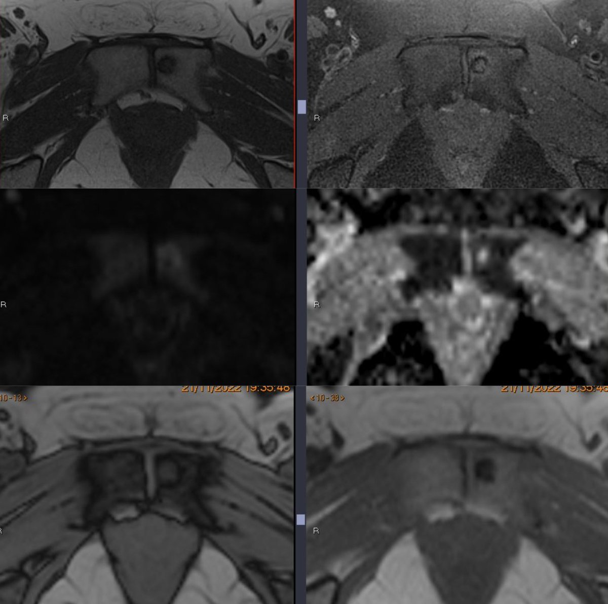
29/8/2023. Some MRI signs "don´t like" bone lesions. History of breast cancer: marrow lesion with sclerotic margins, necrotic center, chemical shift artifact, avid FDG uptake + restricted DWI + peripheral enhancement. CT-guided biopsy confirms metastasis.🔍🦴#Cancer@pepelermarx
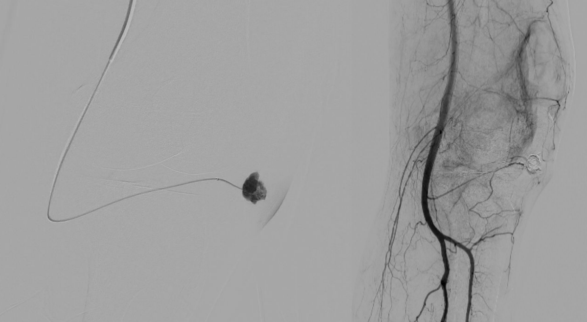
29/8/2023. Explore my latest case: Endovascular geniculate artery embolization – the leading solution for intra-articular pseudoaneurysms causing extensive hemarthrosis, an uncommon complication of knee arthroscopy. #Yinyangsing#Arthroscopy🦵@pepelermarx
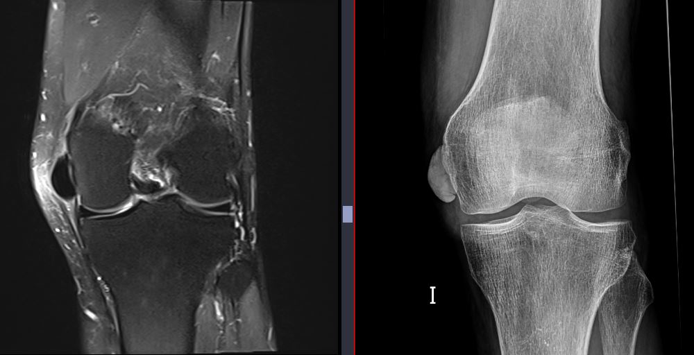
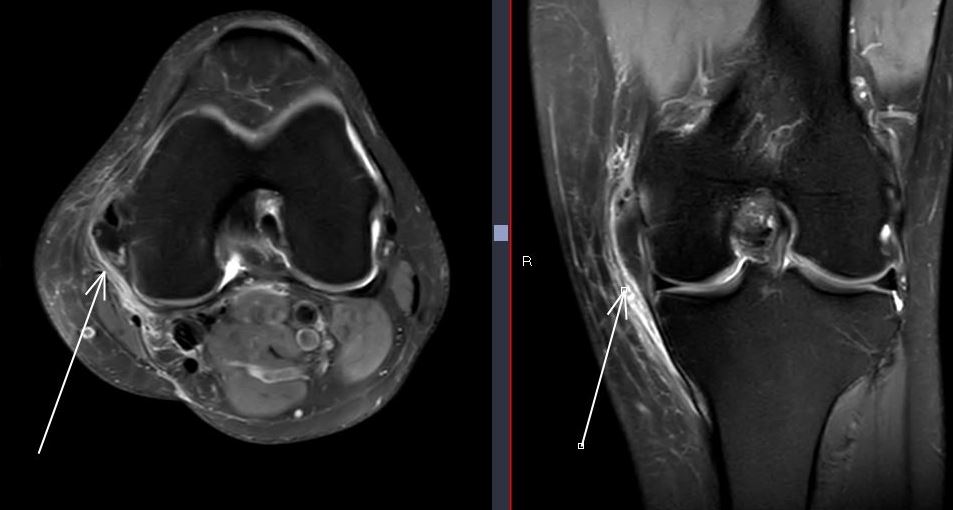
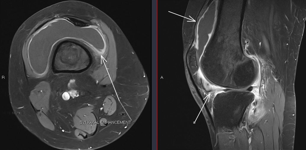
28/8/2023🧩🦵 Another interesting case of calcifying periartritis of the knee, early stage. Type I calcification along the capsular plane of the collateral ligament, minimal pericapsular edema. No anserine bursitis, no bone marrow edema. #MSK@pepelermarx@drwillyjara
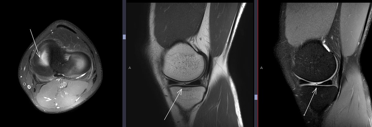
28/8/2023📊🦴 Periarthritis with Type III calcification in the collateral ligament, migrating to the internal epicondyle & anserine bursa. Plus peritendinitis of the pes anserinus. Unveiling the complex world of long-standing knee pain. #MSK#Knee@pepelermarx
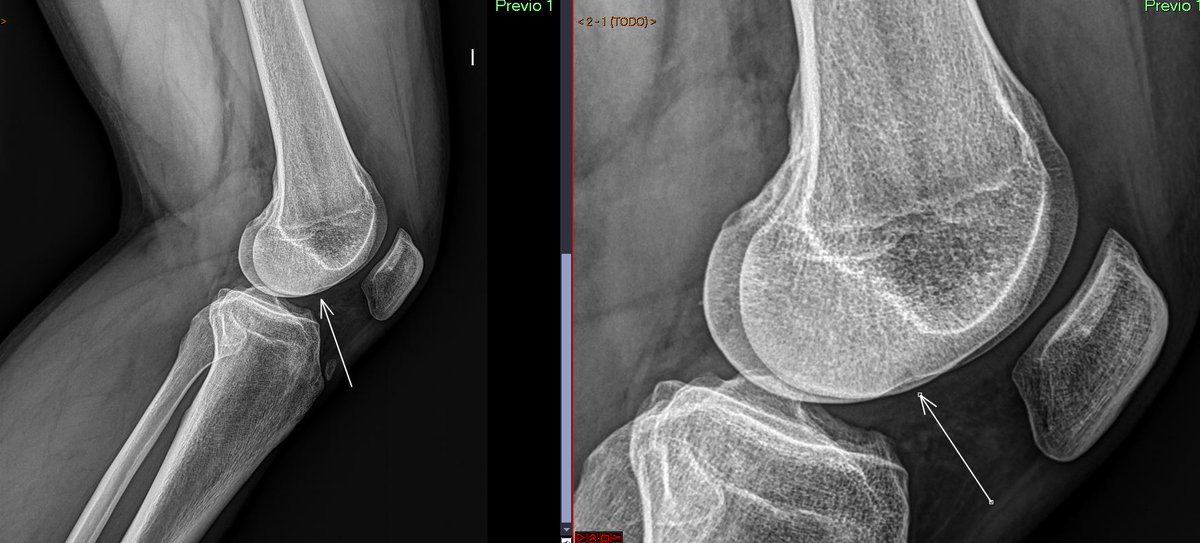
📊 Identifying the "Deep Lateral Femoral Notch Sign" in lateral knee X-ray. This finding may suggest a subchondral fracture, which has been shown to correlate with anterior cruciate ligament tear. 💡🦴 #MSK@pepelermarx@JM_Zulueta_O@drwillyjara
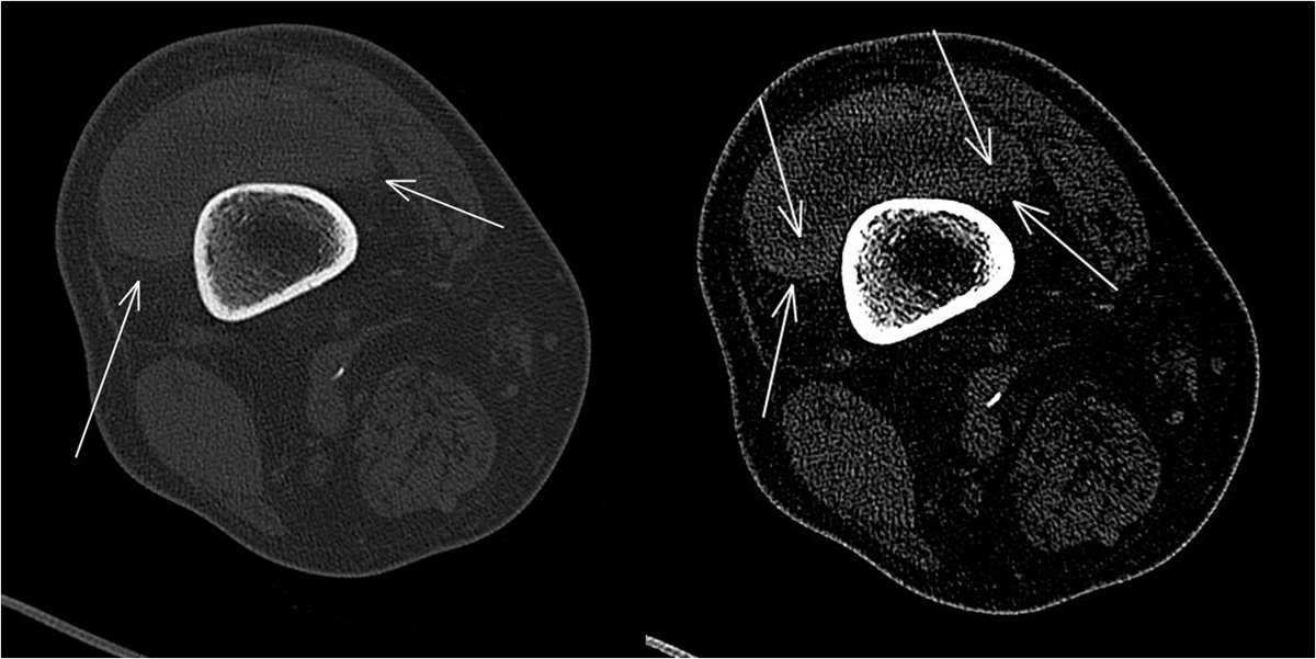
Dual image displaying estimated dimensions of the myofascial defect / muscle hernia.
Check out Radiopaedia for further details on muscular hernias: https://t.co/nxghLD2CXd
#MedEd#MedTwitter#FOAMed#radiology
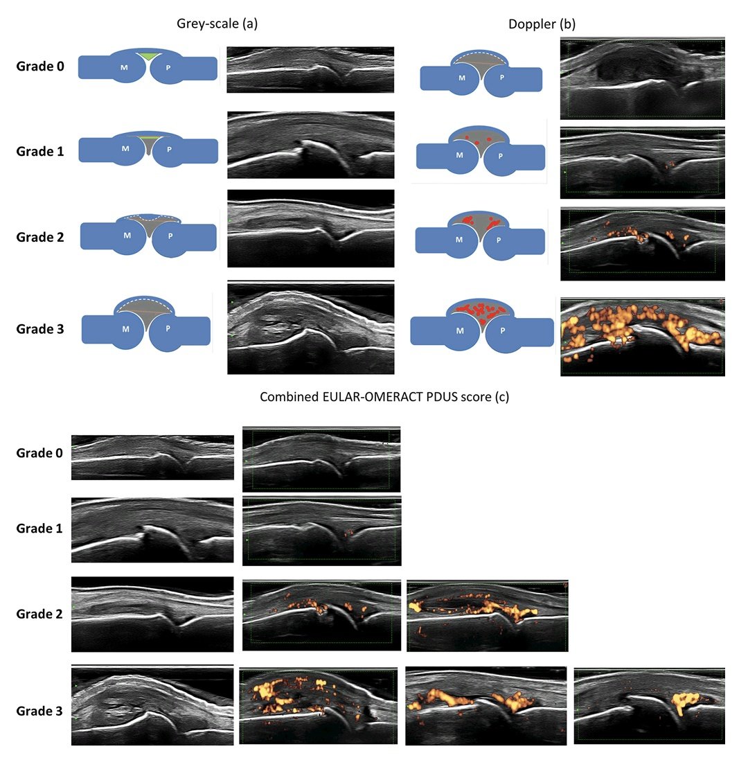
Scoring ultrasound synovitis in rheumatoid arthritis: a EULAROMERACT ultrasound taskforce— Part 1: definition and development of a standardised, consensus-based scoring system
FREE PDF
https://t.co/Kj1cWwFtku