As millennials and gen x, we all chose a side here. The people that we call toxic today as adults started by backing the wrong person during this mess. 😅
Radiology used to have something that was almost invisible because it was so routine:
The reading room consult.
Rounding teams came down.
Surgeons pulled up their cases.
Internists brought the scan that didn't quite fit the clinical picture.
Sometimes the radiologist solved the problem.
Sometimes the clinician supplied one piece of history that completely changed how the images were interpreted.
And usually everyone simply walked away a little sharper.
But something else was happening during all of those conversations.
We were learning each other.
As a radiologist, I learned what individual physicians cared about.
I knew which findings mattered to a particular surgeon.
Which measurements an oncologist was following.
Which details a specialist wanted emphasized.
What questions they were really asking, even when the order just said "pain."
And they learned me.
They knew who was reading their study.
They knew they could walk in, point at something, challenge an interpretation, or ask, "What do you think?"
That relationship created context.
It created feedback.
It created trust.
And over years, it created a kind of institutional knowledge that never appears in the medical record.
When radiology becomes a report produced by someone hundreds or thousands of miles away, we don't just lose proximity.
We risk losing that entire feedback loop.
The report may still be technically excellent.
But the radiologist knows less about the physician.
The physician knows less about the radiologist.
The radiologist sees less of what happened after the report.
And both sides lose opportunities to make each other better.
The reading room was never just a room.
It was where imaging became part of the clinical conversation.
We should think very carefully before designing that conversation out of medicine.
Inspired by a conversation yesterday with @PaleoOnc . Also @909One and @brianchiong .
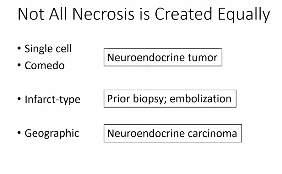
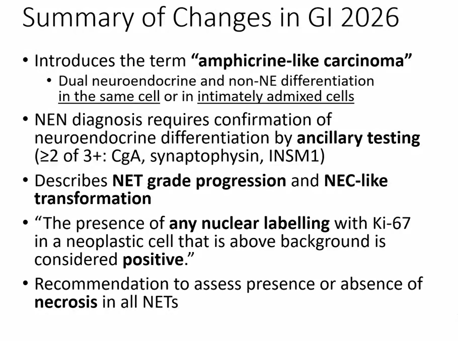
WHAT'S NEW IN NEUROENDOCRINE TUMORS (WHO 6TH EDITION 2026)
Assess presence/abscence of necrosis: Not all necrosis are equal
-Single cell/comedo: NET
-Infarct type: Seen in setting of prior biopsy/embolization
-Geographic: NEC
Dr. Bellizzi GIPS pathcast https://t.co/hVmAzdKI6O #pathology #PathX
WHO 6th ed Gyn path update:
➡️SBT with microinvasion (<5 mm):no longer recommended,describe descriptively.
➡️The term “microinvasive ca” for LGSC like areas <5 mm in SBT is also no longer recommended.Just call it LGSC in SBT😮
This will take some time to get used to!
#gynpath
Updates from the WHO Classification of Breast Tumors, 6th Edition (not published yet)
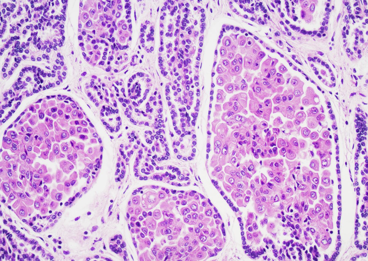
Invasive lobular carcinoma - More detail on special architectural patterns
Solid papillary ILC
• Presence of pseudocapsule
• Often with satellite foci of ILC at the periphery
• Awareness to avoid misclassifying as a papillary lesion especially on small biopsy samples
Dr. Tan #USCAP #pathology #PathX #PathTwitter
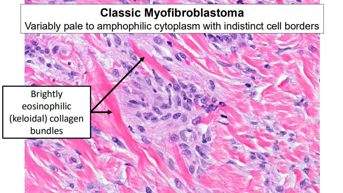
"Common" bland spindle cell lesions of breast
Myofibroblastoma
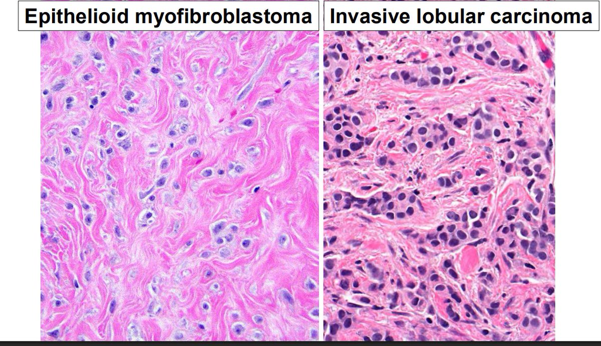
- There are multiple histologic patterns (pic 1)
- Epithelioid myofibroblastoma can be confused histologically with invasive lobular carcinoma.
- Both are ER positive.
- Always run a keratin IHC
Slides from Dr. Cimino-Mathews #USCAP #pathology #PathX #pathtwitter #breastpath
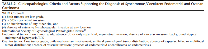
In 2016, two landmark studies showed that synchronous endometrioid endometrial and ovarian carcinomas, often classified as independent primaries, are actually clonal in the majority of cases
Evidence suggests these tumors are of endometrial origin with secondary ovarian involvement
Despite their clonal relationship, prognosis can be excellent when “low-risk” features are present (see pic below)
FIGO 2023 endometrial carcinoma staging now includes substage IA3 for low-risk coexistent endometrial and ovarian carcinoma (CEOC)
If a CEOC does not meet low-risk criteria, FIGO recommends staging it as endometrial carcinoma with spread to the ovary
Rarely, patients have truly unrelated synchronous tumors (e.g., endometrioid endometrial carcinoma + incidental ovarian mucinous borderline tumor/carcinoma). These are staged and managed as independent primaries and are not included in the CEOC category
You can check different CEOC presentations and classification: Huvila et al. Coexistent Endometrial and Ovarian Carcinoma: Terminology, Clinicopathological Features, and Clinical Significance. DOI: 10.1097/PGP.0000000000001177
#GynPath #Pathology #PathX
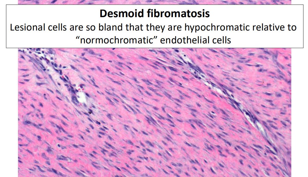
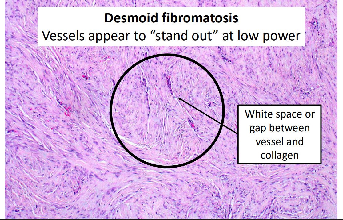
"Common" bland spindle cell lesions of the breast
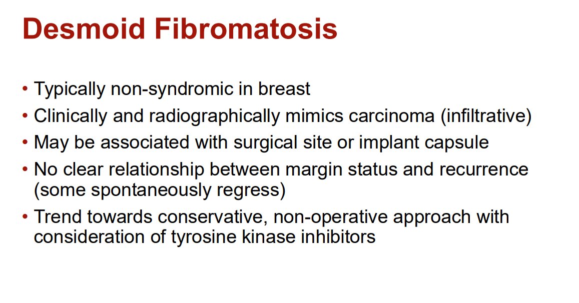
Desmoid Fibromatosis
- Typically non-syndromic in breast
- Clinically and radiographically mimics carcinoma
- Trend towards conservative, non-operative approach with consideration of tyrosine kinase inhibitors
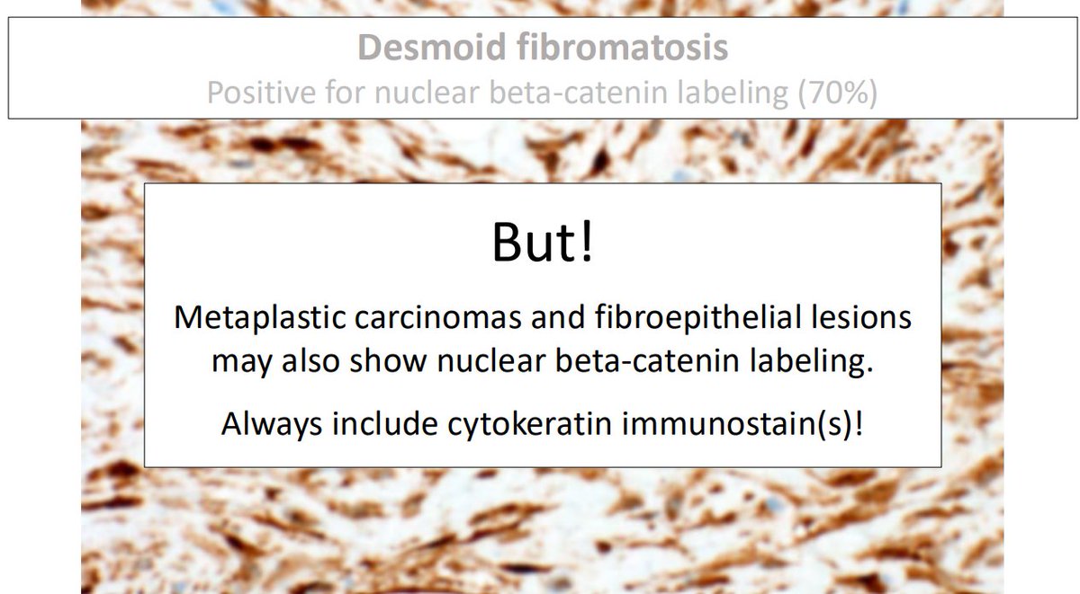
- β-catenin positive (up to 70%), but metaplastic carcinomas and fibroepithelial lesions may also show nuclear β-catenin labeling; always include cytokeratin IHC
Dr. Cimino-Mathews #USCAP #pathology #PathX #PathTwitter #Breastpath
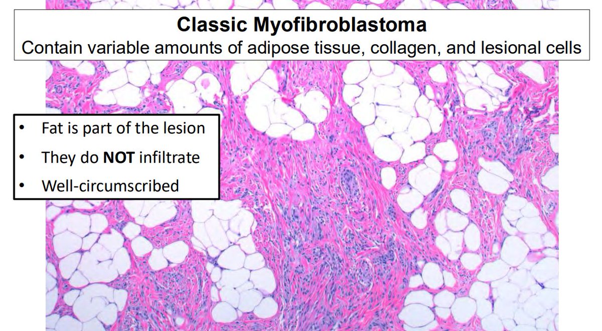
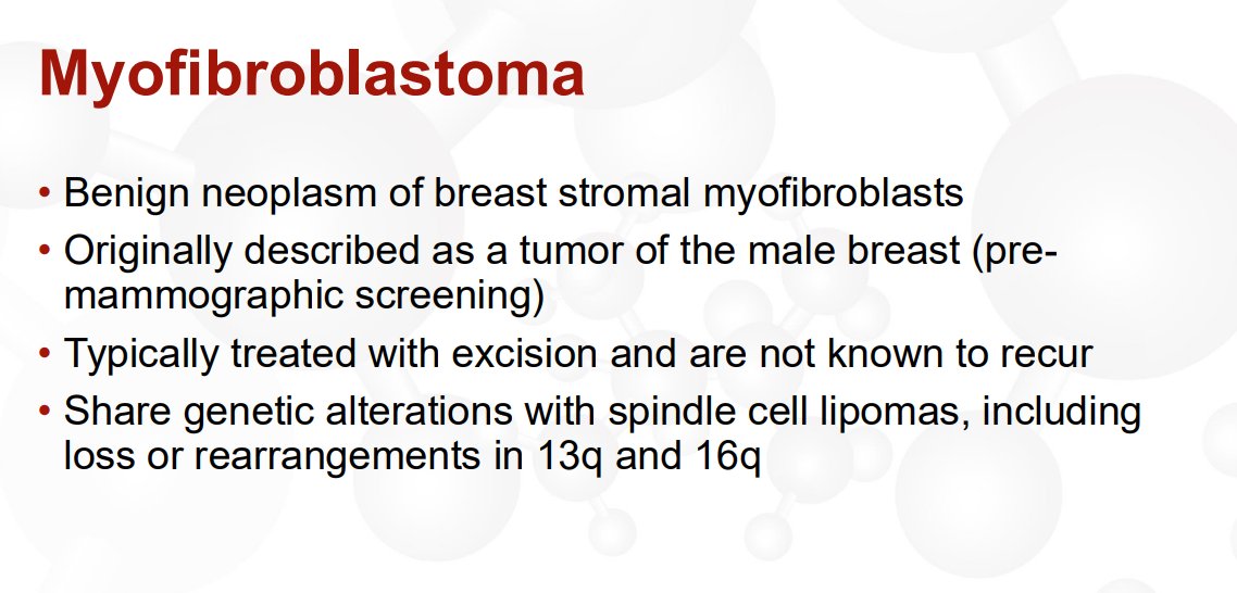
"Common" bland spindle cell lesions of breast
Myofibroblastoma
- Benign neoplasm of breast stromal myofibroblasts
- Typically treated with excision and are not known to recur
- Share genetic alterations with spindle cell lipomas
- IHC: Positive for CD34, ER, SMA and desmin
Dr. Cimino-Mathews #USCAP #pathology #PathX #pathtwitter #breastpath