Thrilled to share Alessandro's beautiful work on a surprising novel presynaptic function of synuclein in dopaminergic neurons! 😎
α-Synuclein and γ-Tubulin Cooperatively Regulate Activity-Evoked Presynaptic Microtubule Nucleation to Gate Dopamine Release https://t.co/CtrJjs6FaM
Our latest collaboration with Guido and Cristina on mechanisms of BIPN! Shared and specific molecular mechanisms of proteasome inhibitors in chemotherapy‐induced peripheral neurotoxicity - Iseppon - British Journal of Pharmacology - Wiley Online Library https://t.co/pxOApYj9Da
Cell biologist Mark Terasaki will give US$25 million of his own money to preserve the legacy of a pioneering scientific institute in Woods Hole, Massachusetts. https://t.co/yutaHoo5pc
A new mechanism for “RNA memory”! 😱
Thrilled to share another crazy paper from the lab (can’t believe we posted 2 in 2 days!), summarizing >10 years of research:
Work on transgenerational inheritance of small RNAs in the powerful model organism C. elegans changed how we think about what’s possible in inheritance and evolution, because it allows the most heretical thing: inheritance of parental responses to the environment! However, it’s still unclear whether RNAs are inherited across generations in other animals, largely because the RNA-dependent RNA polymerases that amplify heritable small RNAs and prevent their dilution in C. elegans are not conserved in mammals.
In this new work, an amazing collaboration with the Rink and Wurtzel labs, we show that planarians establish long-lasting and heritable small RNA–based gene regulatory states despite lacking canonical RNA-dependent RNA polymerases and nuclear RNAi machinery (that are required in C. elegans).
You might say “they are both worms…” BUT planarians are evolutionarily very distant from C. elegans (flatworms vs. roundworms, diverged more than 500 million years ago), making this particularly surprising. These are totally different animals.
We find that ingestion of double-stranded RNA induces sequence-specific silencing that persists for months and survives repeated cycles of whole-body regeneration. Even more strikingly, RNAi can be transferred between animals, echoing James V. McConnell’s controversial “RNA memory” experiments from the 1970s (his lab was targeted by the Unabomber terrorist Ted Kaczynski, who sent McConnell a bomb. This and other controversies ended this line of experiments…)
Mechanistically, we find that the response transitions from a transient systemic dsRNA-triggered phase to a stable, cell-autonomous post-transcriptional “memory phase” maintained by antisense small RNAs. Using a new luminescence reporter (transgenesis is currently impossible in planarians), we show that silencing spreads along the targeted gene and identify a weird type of planarian small RNAs with untemplated polyA tails.
RNAi inheritance without canonical RdRPs establishes planarians as a powerful system for studying RNA-based regulatory inheritance beyond C. elegans and raises the possibility that RNA-mediated inheritance may be more broadly conserved in animals, potentially even in mammals.
Here’s a video of a planarian that is treated by RNAi against β-catenin and develops multiple heads instead of just one. This is one of the phenotypes that is inherited. Another phenotype is “loss of eyes” (which we show is not only inherited across multiple regeneration cycles, but can also be transmitted between animals in transplantation experiments).
Amazing work led by first authors Prakash Cherian and Idit Aviram (co-supervised by Omri and me).
Please read the preprint, the link is in the next tweet, and share!
No-cost workshop opportunities for grad students, postdocs, & trainees! Our Janelia Research Campus is accepting apps for 3 specialized, intensive workshops w/ presentation & networking opportunities. Accommodations, meals, & travel expenses covered: https://t.co/WNKZSp6OgO.
New @CellCellPress paper from Bergles lab at Johns Hopkins just built the most comprehensive map of brain myelin ever made — every oligodendrocyte, across the entire mouse brain, across the lifespan.
The scale: >10 million cells per brain, terabyte-scale 3D lightsheet volumes, registered to the Allen Brain Atlas across 417 regions from 2 months to 2+ years of age.
The technical stack:
Custom tissue clearing (CUBIC-L + SHIELD + uRIMS with 40% urea) to preserve endogenous fluorescence. 3D Mask R-CNN for instance segmentation — not just semantic, instance — so it can distinguish individual cells within dense clusters at scale via overlapping sliding windows. Vision Transformer to classify newly-formed vs. mature oligodendrocytes using soma morphology. All cross-referenced against Allen ISH
transcriptomics and MICrONS serial EM.
What they found:
Oligodendrocyte density varies 10,000-fold across brain regions. Left-right hemispheres: r=0.99. Sex: no significant difference. Strain: matters.
The brain never stops myelinating. New oligodendrocytes are still being generated in 2-year-old mice. Prefrontal cortex L6 shows the fastest rates of new myelination into old age — the circuits for executive
function keep rewiring throughout life.
After demyelination, L4 sensory cortex is the most resilient — oligodendrocytes survive at higher rates. The hippocampus loses nearly everything and barely recovers. Degree of injury doesn't predict rate of
recovery. These are independent axes.
The Alzheimer's result is the most surprising:
Dense-core plaques dominate in cortex and hippocampus. Diffuse/small-core plaques dominate in white matter fiber tracts. Old assumption: diffuse plaques are "less toxic." The data says the opposite — small plaques in fiber tracts cause more myelin loss per plaque than dense-core plaques in gray matter. Plaque load and oligodendrocyte loss are essentially uncorrelated (ρ=0.22). The damage is plaque-type and location specific, not load-dependent.
For MS and AD research: you can't read off white matter injury from gray matter plaque burden. The pathology in fiber tracts is running on different rules.
Data: https://t.co/lIGxb4syZ1
Paper: https://t.co/G5Gsacd4Xw
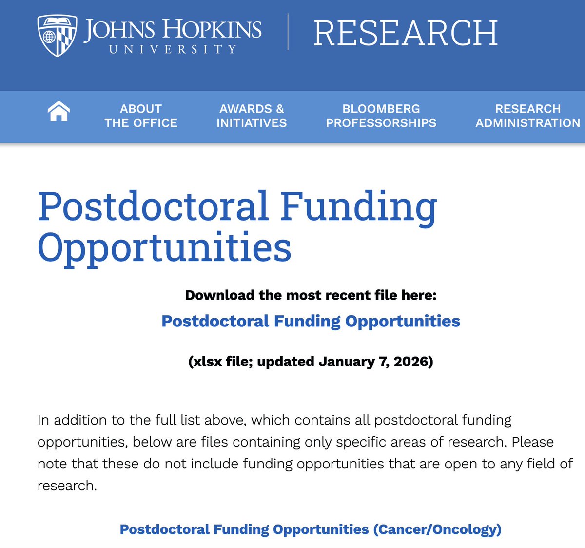
279 postdoctoral fellowships!
Download freely our database of postdoctoral fellowships and grants. For each entry, we provide eligibility criteria, $ amount, deadline, etc.
We also provide separate databases for oncology and neuroX.
Good luck!
Here: https://t.co/EbTahdzbkp
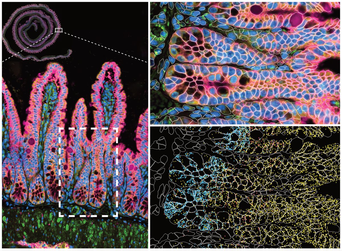
Pain-sensing neurons in the intestines play an important role in defending the body from threats.
New study led by the Artis Lab at @WeillCornell shows TRPV1+ nociceptors in the gut activate tuft cells, leading to expulsion of parasites.
🔗 https://t.co/risDxU1eRs
🚨The Neurocyto lab is branching out in our latest preprint! We used tubulin microinjection to visualize microtubule turnover in developing neurons, demonstrating the presence of in-lattice repair and stabilization in the nascent axon. Check below 🧵1/9
https://t.co/uUDjAixu7y
Wow! So cool🤯 Satellite glial cells can deliver mitochondria to neurons through channels called tunneling nanotubes. Congratulations to Ru-Rong Ji's lab @DukeU! https://t.co/m0ff2U2dDT
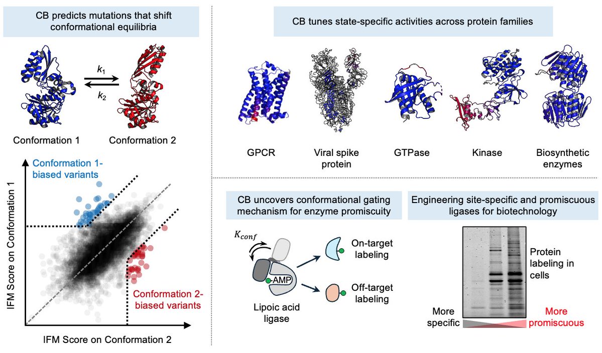
Can we design mutations that predictably bias proteins towards desired conformational states?
Today in @ScienceMagazine, we introduce Conformational Biasing (CB), a simple and scalable computational method that uses contrastive scoring by inverse folding models to identify conformation-biasing mutations.
https://t.co/lbWzHNdMRJ
A tour-de-force circadian proteomics study shows daily rhythms in ~19,000 proteins across 32 tissues in mice, revealing the pervasive reach of the circadian clock. #CircadianBiology#Proteomics#SystemsBiology@MolecularCell
https://t.co/Eom68O7KCB
Today in @Nature we report a new prime editing strategy that can rescue a common cause of many genetic diseases in a disease-agnostic manner. This approach converts a redundant endogenous tRNA into an optimized suppressor tRNA, enabling a single prime edit to rescue premature stop codons across different diseases.
(1/15)
https://t.co/zs0qu5bhXx