☢️ 🍿Radiation-induced cavernous malformations along the cauda equina nerve roots can mimic leptomeningeal disease!
🔷Helpful clues:
1️⃣Negative CSF studies
2️⃣Stability over time
3️⃣Radiation scars
4️⃣Lack of findings outside the radiation field
#Medicine#neurology#radres #neurosurgery #futureradres #Oncology #MRI #FOAMed
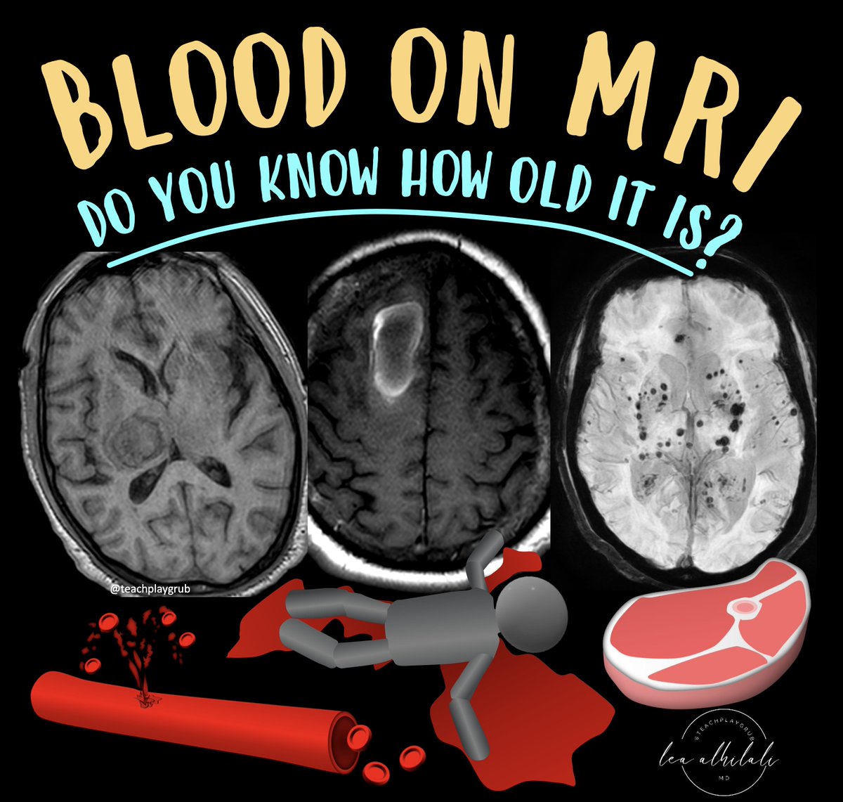
1/Asking “How old are you?” can be dicey—both in real life & on MRI! Do you know how to tell the age of blood on MRI?
Here’s a thread on how to date blood on MRI so that the next time you see a hemorrhage, your guess on when it happened will always be in the right vein!
Epidural hematomas (EDHs) typically do not cross cranial sutures (including midline) due to the firm attachment of the dura to the bones at the sutures.
However, there are exceptions where an EDH can cross suture lines:
1. Fracture-Associated – In cases of diastatic fractures (fractures widening sutures), the epidural hematoma can extend along and beyond the suture.
2. Certain midline locations, such as the vertex or occipital region, EDHs can cross sutures. This occurs because these areas may involve venous bleeding from structures like the superior sagittal sinus, allowing the hematoma to extend across sutural boundaries.
3. Post-Surgical Cases – Following craniotomy or trauma disrupting the dura, EDH may extend beyond normal dural attachment sites.
—pearls and pitfalls from my reporting list
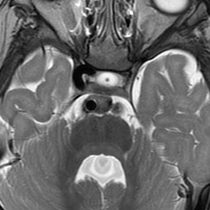
Joubert syndrome is a congenital hindbrain anomaly characterised by a vermian hypoplasia, a midline vermian cleft, thick horizontal superior cerebellar peduncles, and a dysmorphic fourth ventricular roof with loss of the normal fastigium. It's caused by mutations in genes coding for proteins involved in ciliary function (hence its classification as a ciliopathy). Additional abnormalities (e.g., polymicrogyria) are common. This patient also had a hypoplastic left internal carotid artery!
🧵Excited to share our latest publication in Clinical Neuroradiology! 🚀🧠 We show that adding post-contrast susceptibility-weighted imaging (SWI+C) significantly enhances the detection of active lesions in multiple sclerosis (MS). (1/5)
1/Does PTERYGOPALATINE FOSSA anatomy feel as confusing as its spelling?
Does it seem to have as many openings as letters in its name?
Are you pterrified of the pterygopalatine fossa (PPF)?
Let this thread on PPF anatomy help you out.
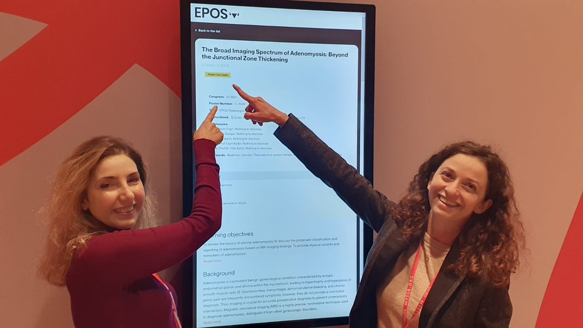
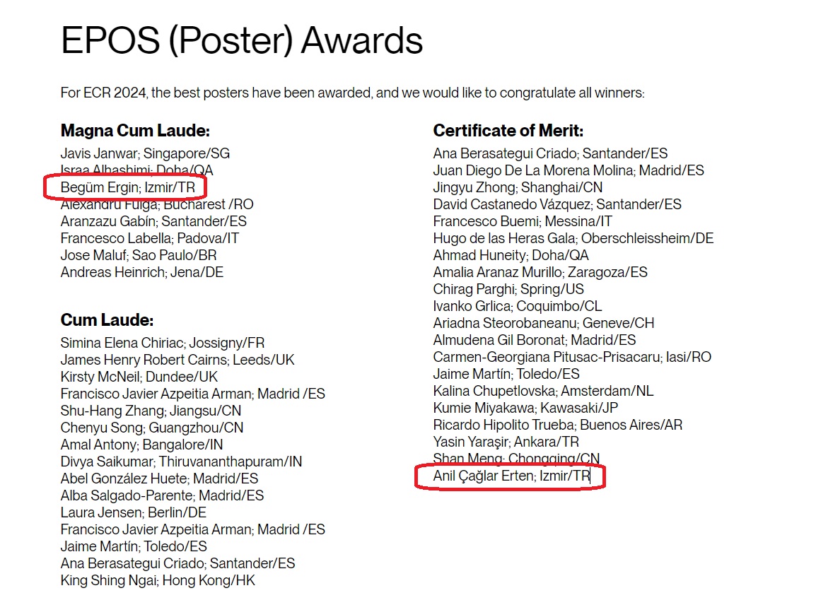
Thrilled to receive the Certificate of Merit Award at #RSNA2024! 🎉 Grateful for the guidance and support of my mentor @YPekcevik whose expertise and encouragement have been invaluable. This is a shared achievement! 🌟 #Radiology#RSNA24
“Ready to tackle salivary gland tumor mysteries at #RSNA24 !🕵️♂️🔍”
Huge thanks to my mentor @YPekcevik , for turning the diagnostic puzzles into ‘piece of cake’ moments. Let’s solve these riddles together! 🧩 @RSNA#RSNA2024
1/Is remembering cerebellar anatomy making you dizzy?
Need help telling your flocculus from your nodule?
How much cerebellar anatomy do YOU know?
Here’s some help w/an anatomy thread on the 9 lobules of the vermis!
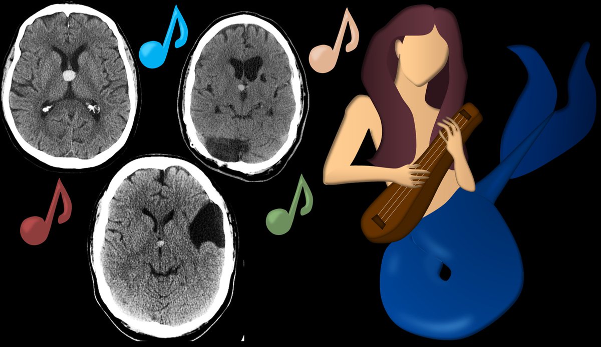
1/Don't fall for the siren song of calling all bright round objects at the foramen of Monro colloid cysts!
Like a true siren song, this may be a TRAP!
If you hear the call of colloid��read this first
Here's a thread about lesions here that can trap you--& how to avoid them...