Who knew amyloid (yes, as in Alzheimer's disease) makes deliveries of RNA to sperm for translation in the embryo?!
@MolecularCell
https://t.co/n3jx84RTyB
How does the mitotic spindle adapt to genomes that differ in size by more than 10,000-fold? Our new paper in @NatureCellBio reveals a simple scaling law and the physics behind it: chromosome crowding! 🧵
https://t.co/DsGvz8ulME
An open model you can chat with about raw DNA sequences. 🤖 Great for access. But sequence is only half the story: biology happens in 3D, in whole bodies. Next wave of bio-AI must learn from where cells and molecules actually are.
https://t.co/6DOvwwCN0C
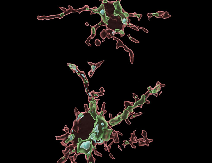
Glia derives from the greek word for glue, but they are so much than that.
In this video by our #ElectronMicroscopy team, shows a pyramidal neuron (PyC) surrounded by 8 glia brain cells – 5 microglia (MG) and 3 oligodendrocyte progenitor cells (OPC).
🔗 https://t.co/dJnUUU9MZx
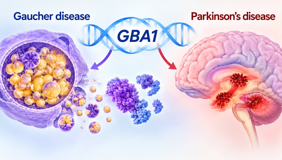
Gan-Or traces the history of GBA1 research from the first description of Gaucher disease to current understanding of how GBA1 drives Parkinson’s disease risk, while examining emerging targeted therapies and future research directions. https://t.co/zqMm80PkPV
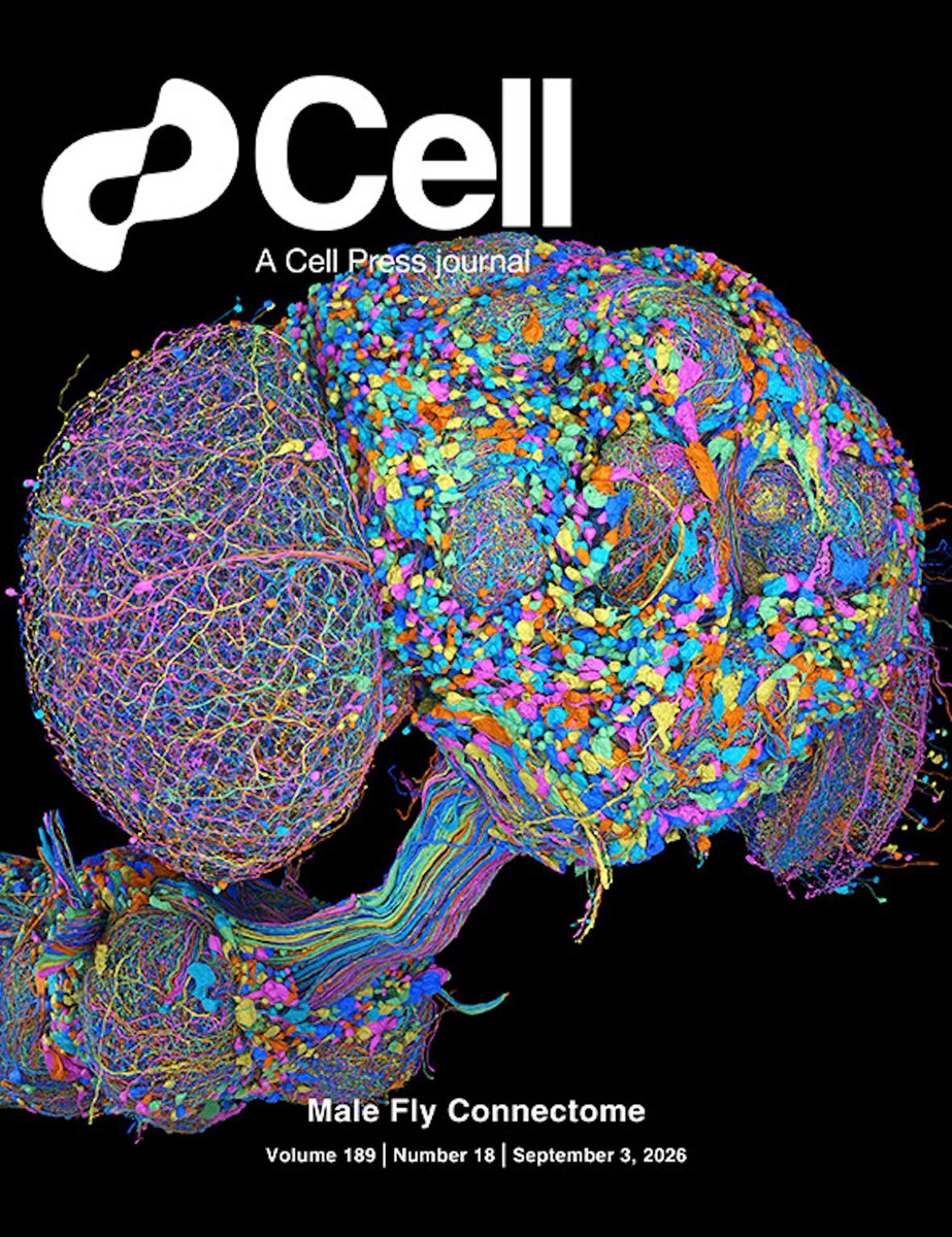
Sexual dimorphism in the complete Drosophila male central nervous system connectome.
Learn more in Cell: https://t.co/1zylFILm1G
Gregory S.X.E. Jefferis & colleagues
@cellcellpress
New issue is out👉https://t.co/uP2kI6DjtL
On the cover: the complete connectome of the male adult D. melanogaster central nervous system. The cover depicts a single representative cell from each of the 11,710 neuron types in the dataset, which contains 166,700 neurons in total.
🌱 2025 was the 1st year of the Seeding Neuroscience Grants program. The response from emerging #neuroscience societies & thematic networks was incredibly positive!
📖 The 2026 call opens in 3 days. Find out more: https://t.co/8paKL1K37l
@ElfarrashSara@Kavita_Babu_@FZolessi
Researchers used a technique that produced a detailed protein map of the human retina. Fluorescent antibodies highlight specific structures in this image captured with #microscopy from Leica Microsystems. Read more: https://t.co/c9Bf0Ch1X7
@BPoD_s@BiochemOxford
This 'important' Tools and Resources paper establishes a robust live-imaging toolkit to characterize excitatory and inhibitory synaptic dynamics during neuronal development. The methodology and quantitative data are 'convincing' and well validated.
https://t.co/24OcZ35AI2
How does white matter recover after #stroke?
A team shows how microglial A-2A receptor repairs white matter by stimulating #efferocytosis and oligondendrocyte differentiation—and can be targeted with micelles to promote recovery in mice. @FudanUniversity https://t.co/1gXkhXaESG
Video of myosin proteins "walking" along actin. Insane that we can image this!
It was made by Toshio Ando's group using atomic force microscopy. All of their papers are a goldmine; they've also recorded a Cas9 protein cutting DNA and ATP synthase spinning.
Our paper "Highly attenuated dendritic propagation of isolated synaptic potentials in vivo", demonstrating that individual synaptic potentials in vivo do not reach the soma, is published in Science Advances: https://t.co/8w0Bz8cUEb