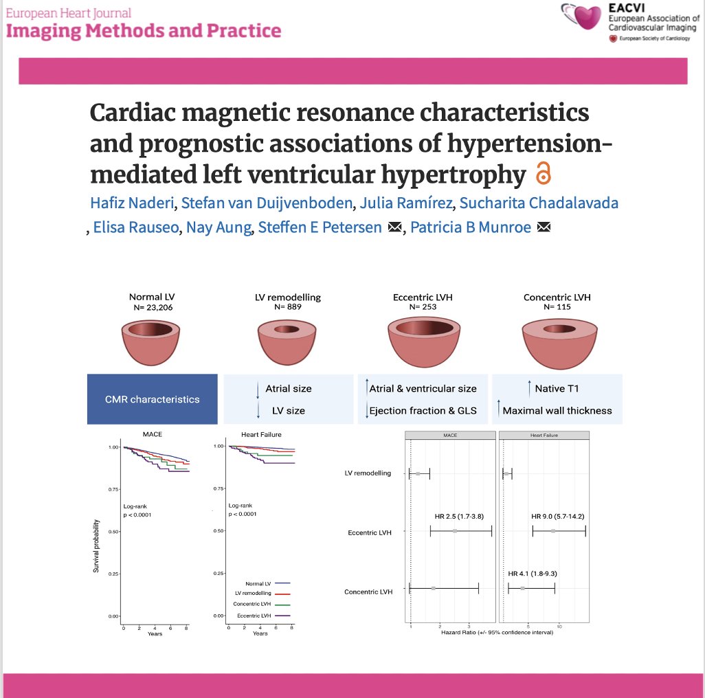
📄 Hypertension-related LVH: not all phenotypes carry the same risk
🔗 DOI: https://t.co/W1SFC0ZCz6
🫀 Hypertension is the most common cause of LV hypertrophy—but its cardiac expression is far from uniform.
This large UK Biobank CMR study (n >24,000) provides a comprehensive look at LVH phenotypes and their prognostic impact.
✨ Four CMR-defined phenotypes:
👉 Normal LV
👉 LV remodelling
👉 Eccentric LVH
👉 Concentric LVH
📊 As shown in the graphical abstract (page 2):
➡️ each phenotype has distinct structural and functional signatures
✨ Key findings:
🔹 Eccentric LVH = worst phenotype
➡️ Most impaired LV function (EF + strain)
➡️ Largest chambers
➡️ Highest risk:
MACE → HR 2.5
Heart failure → HR 9.0
🔹 Concentric LVH:
➡️ Highest wall thickness and native T1 (fibrosis)
➡️ ↑ Heart failure risk (HR 4.1)
➡️ No significant MACE association
🔹 LV remodelling:
➡️ Intermediate phenotype
➡️ Smaller chambers, milder changes
📊 Key pathophysiological insight:
👉 LVH is not a binary condition—but a spectrum of myocardial adaptation
➡️ From remodelling → concentric or eccentric hypertrophy
➡️ Driven by pressure load, volume load, and myocardial response
💡 Clinical take-home message:
👉 Not all LVH is equal
✔ Eccentric LVH → high-risk phenotype
✔ Concentric LVH → fibrotic, HF-prone phenotype
👉 CMR enables:
precise phenotyping
improved risk stratification
potential tailored treatment strategies
🚨 Bottom line:
In hypertension, LV geometry matters—because different phenotypes carry very different prognoses.
#Cardiology #CMR #Hypertension #LVH #CardiacImaging #HeartFailure #RiskStratification #PrecisionMedicine #UKBiobank 🫀📊
1/10
Coronary artery calcium (CAC) scoring is a quick CT scan that detects calcified plaque in your heart arteries — often called a powerful tool for predicting heart attack risk.
But large studies show the added value is more limited than the hype suggests.
Here's what the evidence (MESA, DANCAVAS, CONFIRM & more) actually says 🧵 #HeartHealth #CACScore
A bright surprise within the heart,
Fat-signal high on every chart—
T1 weighted, shining bright,
Suppressed by fat-sat, out of sight.
Encapsulated, smooth, benign,
No enhancement post-contrast line.
Silent guest in cardiac space,
A lipoma in its gentle place.🫀✨
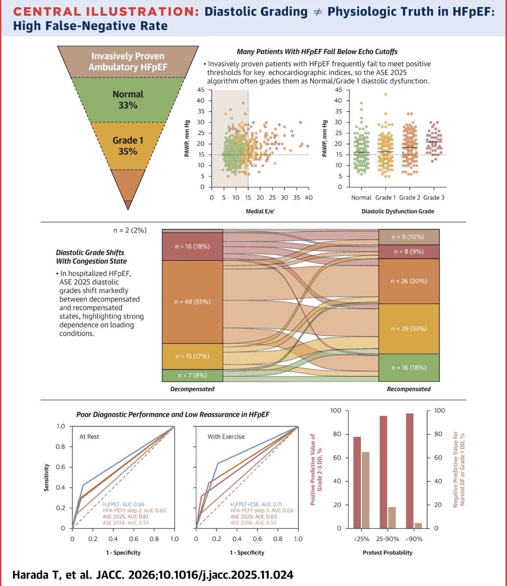
Echo DD guidelines are consensus based and require testing to determine clinical utility. In this multicenter study with gold standard dx, we show poor sensitivity and accuracy for resting and exercise echo criteria. @JACCJournals
https://t.co/x64Fl4Sh3X
1-Fenestrations of aortic valve .Normal or Abnormal?
History
Rokitansky gave an accurate account of valvular fenestration, a condition which he regarded as a form of atrophy. Very little can be added to his original description which follows:“These perforations are almost always
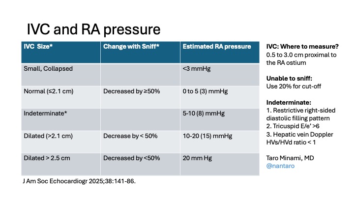
Based on the new ASE guideline (J Am Soc Echocardiogr 2025;38:141–86), I put together this concise IVC–RA pressure table. Hope this is helpful. #POCUS#IVC
Calculation of Regurgitant Volume Using #echofirst Volumetric Method for Accurate Diagnosis of Severe Mitral Regurgitation - Journal of the American Society of Echocardiography https://t.co/Kw3Ena6zve
1️⃣
Some patients with severe venous congestion have almost no oedema — and that’s confusing at first.
It only starts to make sense once you unpack the physiology. 👇
🧵 Starling’s Law: Misunderstood, Misapplied, and Still Misleading
1
🚨 “Starling’s Law explains how the heart increases cardiac output.”
You’ve probably heard this a thousand times.
But it’s wrong.
Or at least - very incomplete.
Let’s fix it.
Because this matters - for heart failure, fluids, vasopressors, inotropes, afterload, and how we think about the whole system.
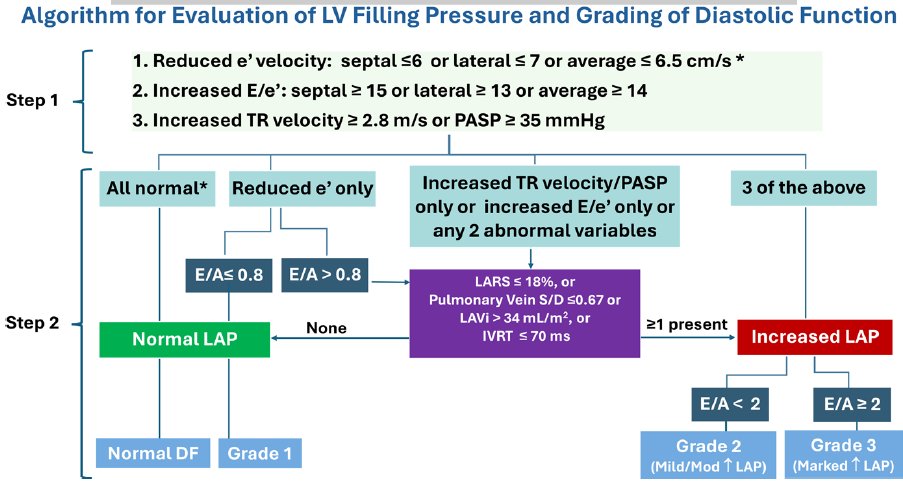
📢New Algorithm for Estimating LV Filling Pressure by Echo:
🔴increase in the number of patients in whom LV filling
pressure can be estimated
🟢only 2 cases of indeterminate LV filling pressure and the majority had a definitive LV filling pattern
https://t.co/4yL751yFFo
Prediction of ventricular arrhythmic outcomes in suspected cardiac sarcoidosis: a comparison of cardiovascular magnetic resonance phenotyping vs. societal recommendations for implantable cardioverter-defibrillator placement
https://t.co/frywfouKi2
#WhyCMR#Epeeps#cardiotwitter
Mitral valve comes with different shapes and forms. A patient with mild to moderate mitral regurgitation. 3D TEE, LA perspective. How would you describe this valve morphology?