After a little rest of our team, we are happy to announce that the Chapter start it’s activities for the 2023 with an astonishing speaker, @hERGologie. Save the date, see you soon 🫡🫡 @bps_students@SOBLA_BIOF@BiophysicalSoc
My lab in Chile kicked off in July. Faced with modest funding and the steep costs of reagents compared to Europe or the USA, we had a thought: Why not create our own molecular biology reagents and kits? This approach isn't just cost-effective; It also serves as a teaching tool!
Electrical signaling in Venus Flytrap shown in real-time⚡️ Touching the trigger hairs of the trap causes an influx of calcium ions (shown on top) that spreads rapidly across the leaf. Two action potentials and the leaves quickly close to trap their prey 🪰
optoKANK is a new optogenetic tool to disassemble individual focal adhesions! 🔦 In the preprint the authors find a local burst of acto-myosin contractility is necessary to disassemble FAs. The burst is caused by GEF-H1 released from microtubules.
https://t.co/pXCMSbfg6b
Un nuevo año buscando científicxs que quieran hacer su postdoctorado en nuestro lab a través del financiamiento de ANID. Si conocen de alguien que quiera realizar su investigación postdoctoral en biofísica e ingeniería de proteínas, pasen el dato! https://t.co/UyslL1vIgT
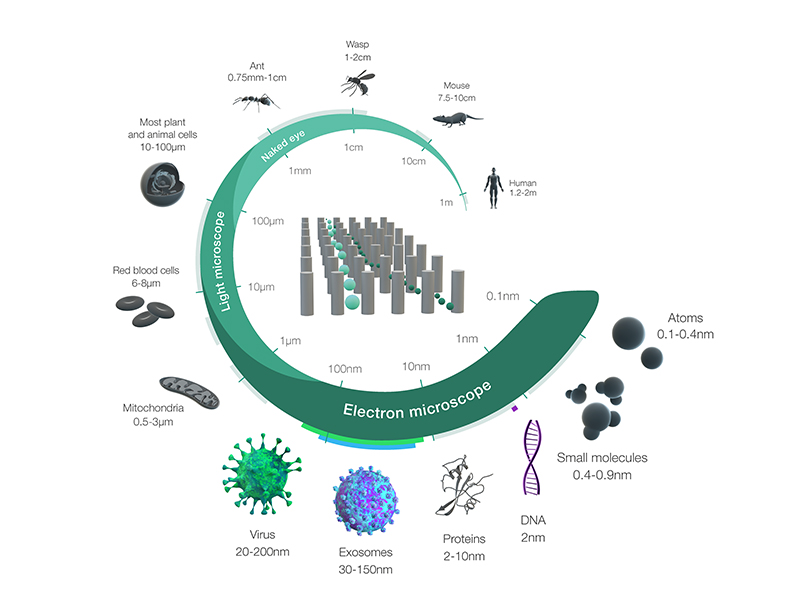
Tips for designing figures for your manuscript! #sciart
1) Keep it simple: The figure should clearly and concisely convey the information you are trying to present. Avoid clutter and unnecessary detail that can distract from the main message.
2) Use appropriate software: Choose software that is suitable for your type of figure. For example, use vector graphics software like Adobe Illustrator for creating diagrams and line drawings, and use graphing software like Excel or Origin for creating graphs.
3) Label everything: Ensure that all labels, axes, and other text are clear and easy to read. Use a font size that is appropriate for the size of the figure, and avoid using too many colors or fonts that are difficult to read.
4) Choose appropriate colors: Use colors that are easy on the eyes and that are easily distinguishable from each other. Consider color-blindness and choose colors that are accessible to all readers.
5) Use high-quality images: Use high-quality images with a high resolution. This will ensure that the details of the image are clear and crisp.
6) Get feedback: Share your figures with colleagues and ask for feedback. This can help identify potential issues or areas for improvement that you may have missed.
7) Follow journal guidelines: Follow the guidelines of the journal you are submitting to regarding figure format, size, and resolution. This will ensure that your figures are presented in the best possible way and meet the requirements of the journal.
8) Keep a record of your figures: Keep a record of all the figures you create, including any changes you make during the editing process. This will help you keep track of the different versions of your figures and ensure that you use the correct version when submitting your manuscript.
9) Be consistent: Ensure that all figures are consistent in terms of style, font, and size. This will help to create a cohesive and professional-looking manuscript.
If you're interested in mechanosensitive ion channels, surface chemistry, or instrument design, check out our recent paper:
'Freestanding lipid bilayer tensiometer for the study of mechanosensitive ion channels' |https://t.co/QUqQosttmu
The miniGFPs plasmids are now available from @Addgene https://t.co/5Bf6rqahqQ
Here is the original manuscript reporting the development and characterization of the smallest GFPs https://t.co/e7co0PvrHB
Forgot to mention at the end of my talk that I’m looking for grad students/postdocs/res scientists curious about how the pacemaker rate adapts. If you are interested let me know!!!
After a little rest of our team, we are happy to announce that the Chapter start it’s activities for the 2023 with an astonishing speaker, @hERGologie. Save the date, see you soon 🫡🫡 @bps_students@SOBLA_BIOF@BiophysicalSoc
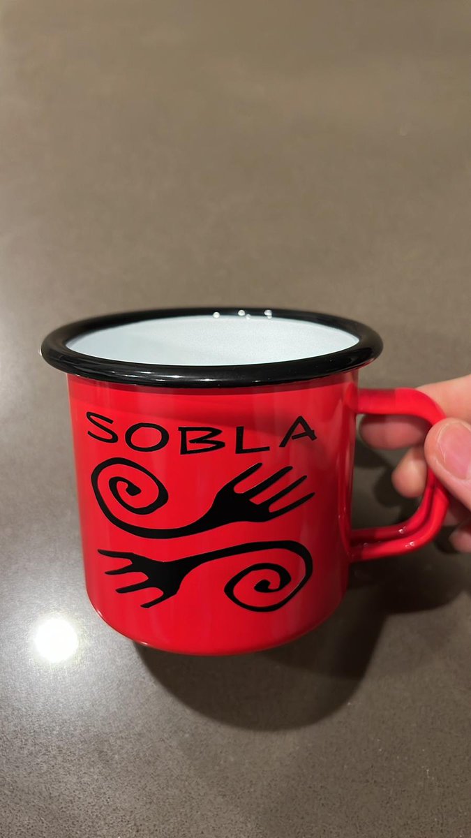
Hello #Twitter! Looking forward our Annual meeting at the @BiophysicalSoc meeting in San Diego! Help us keep up the good work 💪by supporting our society & getting one of these ⚡️wonderful coffee mugs!Is red❤️ you color?We have many more🌈! Get them at https://t.co/DFwegI1QUF😍!
Our latest preprint💥 Tissue deformation is critical for organ generation, but how do complex organs consisting of multiple compartments and cell types achieve this? A fantastic interdisciplinary collab with @ManningResearch https://t.co/zdBK65cqO9 short🧵👇