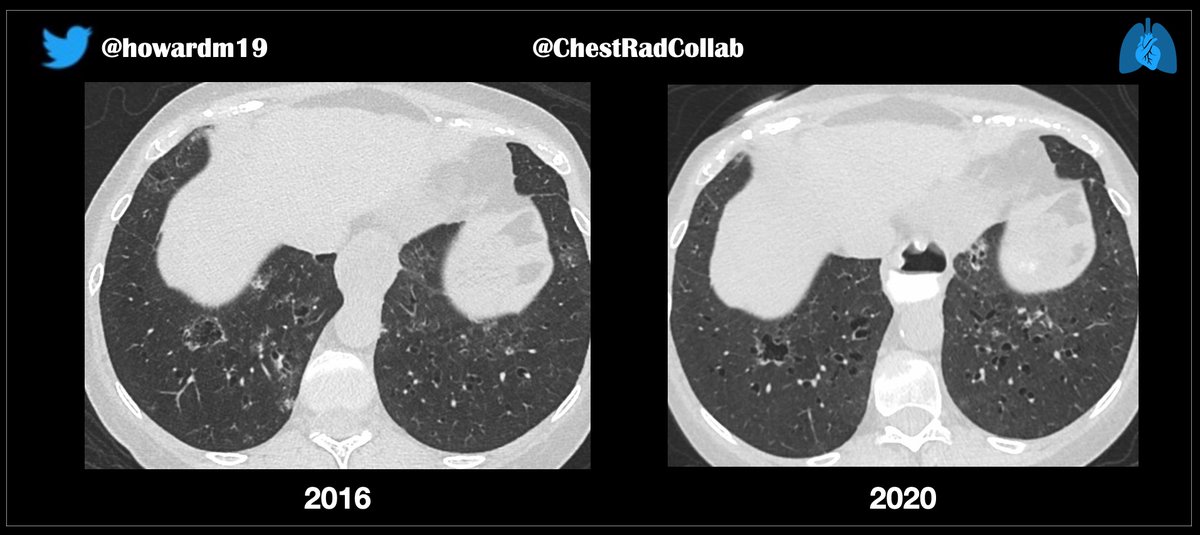
@JeffreyKanneMD @TanMohammedMD @LucyModahlMD@smlungpathguy @LungPathGuys
Case for STR Webinar tomorrow. (3 images)
Patient with Sjogren syndrome & limited cutaneous scleroderma.
• what are the findings ?
• by what pathomechanisms are they formed ?
#pulmpath#ChestRadED
@howardm19 @JeffreyKanneMD @TanMohammedMD @LucyModahlMD@smlungpathguy @LungPathGuys 1. Gottron papules and Mechanic's Hands
2. I was anticipating an anti-synthetase Ab. But, no, it was:
3. Anti-MDA-5 Ab.
That's not good. This Ab. may be associated with bad lung disease. The 2nd CT shows progression with basal fibrotic NSIP pattern.
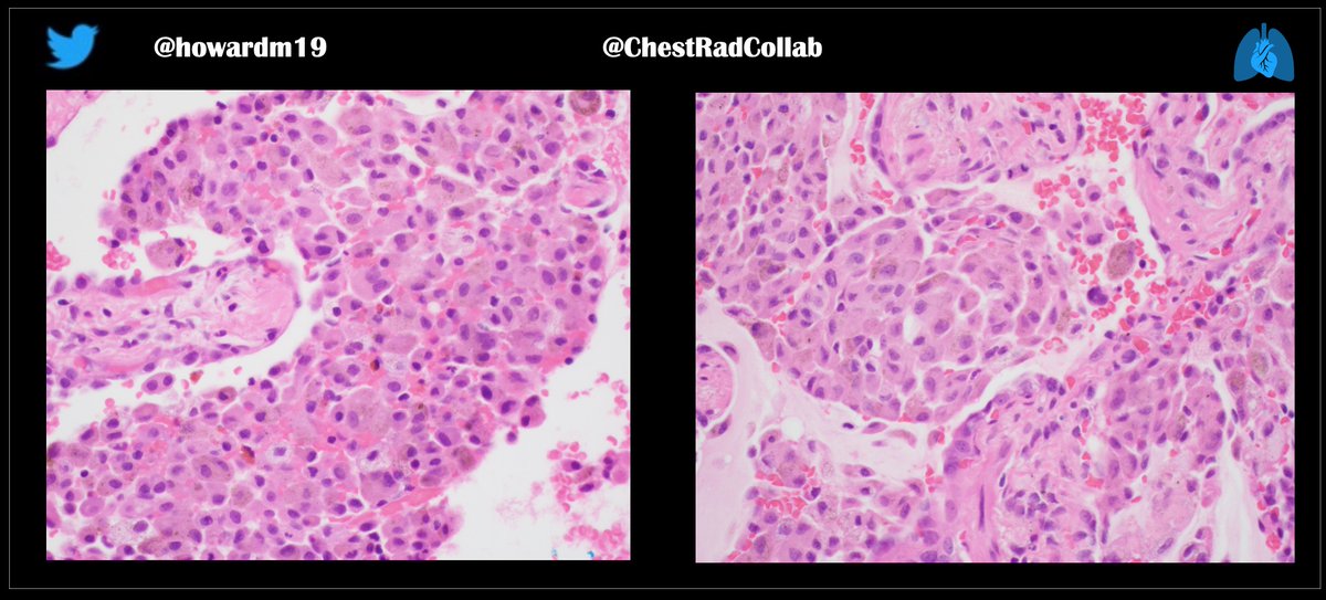
@smlungpathguy@6Patterns @JeffreyKanneMD @yro854
A 60-year-old female with a chronic respiratory illness. A surgical lung biopsy was performed. History withheld.
(See all three images below).
Diagnosis ?
#ChestRadEd#FOAMrad
@yro854@smlungpathguy@6Patterns @JeffreyKanneMD Indeed. She is a smoker.
The massive accumulation of smoker’s macrophages produce the diffuse pulmonary hyper-attenuation abnormality.
I (non-pathologist) think the appearance of the pigment is typical
On CT, I do not see findings of interstitial fibrosis, as such.
@howardm19 @JeffreyKanneMD
Blunt chest trauma. Acute aortic mural injury.
Have you ever seen this pattern of injury — see white arrow— before? Abdominal scan acquired minimally later as indicated by time stamp.
Retweet to your colleagues.
Comments and images are welcome.
@howardm19 @JeffreyKanneMD This is the so-called “neovascularity” of severe pulmonary hypertension.
1. Tortuous/corkscrew vessels
2. Ground-glass opacities, often in relation to the vessels.
These are not AVMs.
This article explicates the pathology:
PMID:16267251
Another: PMID: 22205444
#ChestRadED
@howardm19 @JeffreyKanneMD
Case for STR Webinar tomorrow.
Patient with severe familial (BMPR2-related mutation) pulmonary hypertension.
How would you describe/report the vessels ? What pathology do you infer ?
#ChestRadEd
@howardm19 @JeffreyKanneMD @howardm19
Yip, I would have diagnosed a LV thrombus, too. :-)
But, the Case of the Disappearing Thrombus…on the portal venous phase.
Nice, BL.
(Case courtesy of DR. Thanks!)
@howardm19 @JeffreyKanneMD
Super Bowl Sunday case. Let’s chat.
Patient with an abdominal neuroendocrine tumor. Surveillance imaging over years. How would you report the findings on the 2019 images ?
#ChestRadEd
@howardm19 @JeffreyKanneMD Pathologic findings: pleuroparenchymal fibroelastosis (PPFE).
It’s usually diagnosed in the context of lung or stem cell transplantation (late complication), and in familial fibrosis (sometimes with an identifiable telomeropathy). That’s not the case in this instance.
@howardm19 @JeffreyKanneMD
STR webinar tomorrow
A surgical lung biopsy was performed for a “suspicion of ILD.”
A geologist for ~ 15 years, sometimes working at mining sites. No details about the type of mining. No family history of ILD.
What’s a likely pathologic diagnosis ?
@howardm19
Answer: Marfan Syndrome. The aortic root is dilated.
A paternal uncle was affected.
No “fancy” genetic testing is necessary.
At https://t.co/KxIRh1I3fl:
#ChestRadEd
@JeffreyKanneMD @howardm19 @TanMohammedMD @mdmq
Case for Webinar tomorrow:
https://t.co/G9Cbn9TTVU
I was presented with “nodule follow-up" case yesterday. Nodule found on calcium-scoring CT.
I said: “Cute” — because my daughters sometimes say that.
#ChestRadEd#FOAMrad
@JeffreyKanneMD @howardm19 @TanMohammedMD @mdmq
Case for Webinar tomorrow:
https://t.co/G9Cbn9TTVU
History of AML; underwent allogeneic SCT in November 2016. It’s not clear why chest CT (below) was performed, but interesting findings are present.
#ChestRadEd#FOAMrad
@i_hate_nodules
@howardm19
@TanMohammedMD
@mdmq
@DarelHeitkamp@StefanTigges
76 M with progressive dyspnea after starting daptomycin for soft tissue infection. What pattern of lung injury? Case to be presented tomorrow by @JeffreyKanneMD
#ChestRadEd#FoamRad#RadRes