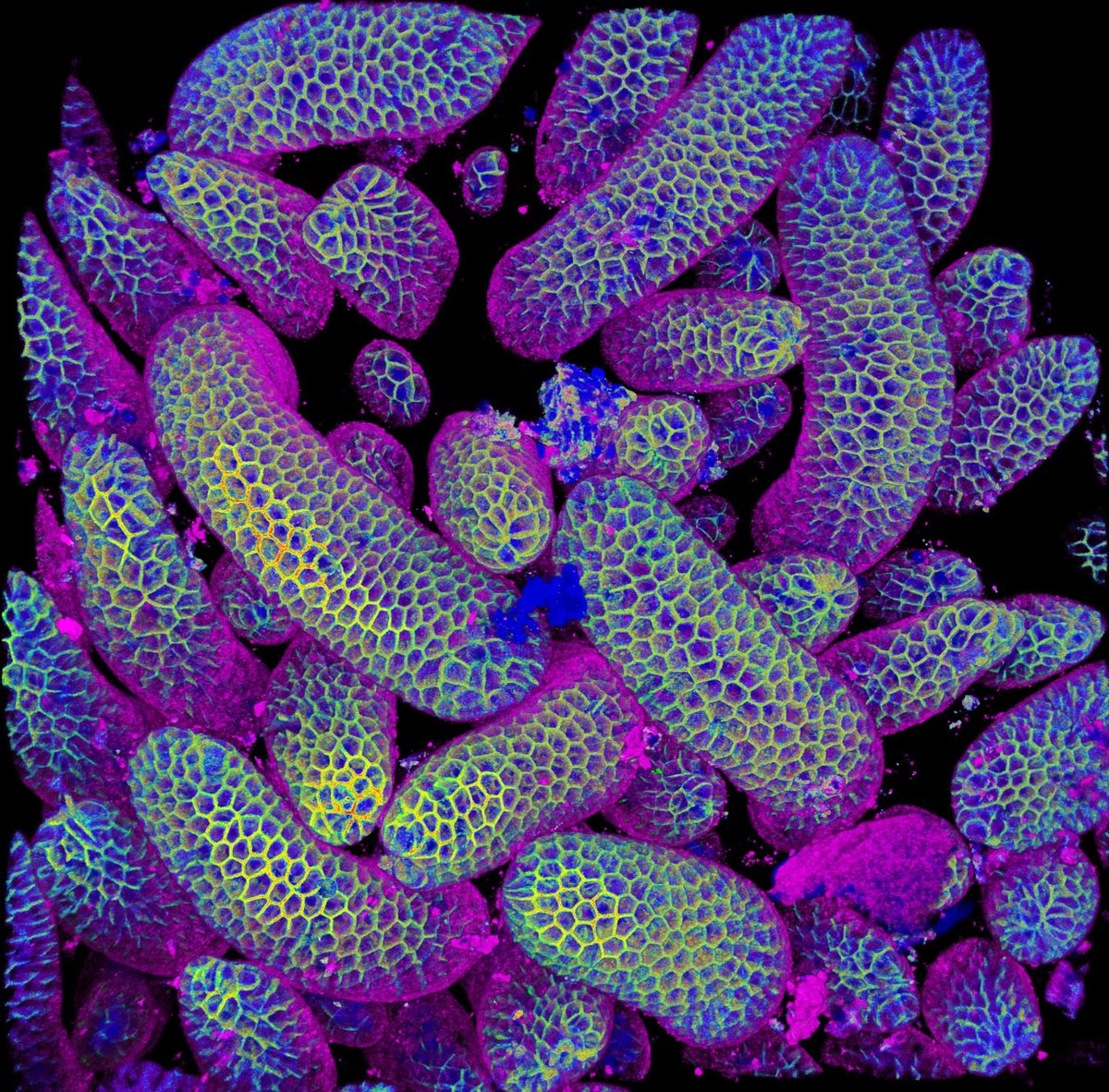
¡Es un honor haber obtenido el 1er y el 20° lugar del concurso de #NikonSmallWorld en su 50° aniversario! 🤩 Es un privilegio mostrar la belleza y la complejidad del mundo microscópico. ¡Gracias @NikonSmallWorld! 🔬🇨🇱
Congratulations to all the winners of our milestone 50th anniversary #NikonSmallWorld competition! We're so grateful for every single person who submitted and made this competition into what it is today. The complete 2024 winning gallery is now live: https://t.co/I8Pcou7949 🔬
ICYMI - we're hosting a new quarterly webinar series on cell migration!
In our first webinar on Thursday 11 June at 15:00 BST (UTC+1), we'll hear talks from Juan Manuel Garcia Arcos, Yohalie Kalukula & Daniel J. Cohen.
https://t.co/DGeEDK2ivF
Microscopy Image Competition 2025 Finalists
There are two categories:
Category 1: Featuring Proteintech antibodies
Category 2: Featuring any antibodies
The images below are the Category 2 finalists.
Vote before Oct 26 (T&Cs apply): https://t.co/vc7YGK3nQx
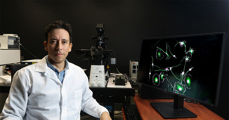
🔬 Meet the researcher behind the image that won the BioTechniques Image Competition 2025 📸
In this interview, we learn more about Bruno Cisterna, his winning image, his favorite microscopy techniques and his advice for burgeoning microscopists >>> https://t.co/x9dMnBi8Ip
🥁 The winner of the 2025 BioTechniques Image Competition is… https://t.co/mRZnPpdY04
Keep an eye out for our follow-up interview with Bruno Cisterna (Medical College of Georgia at Augusta University) to learn more about this beautiful image!
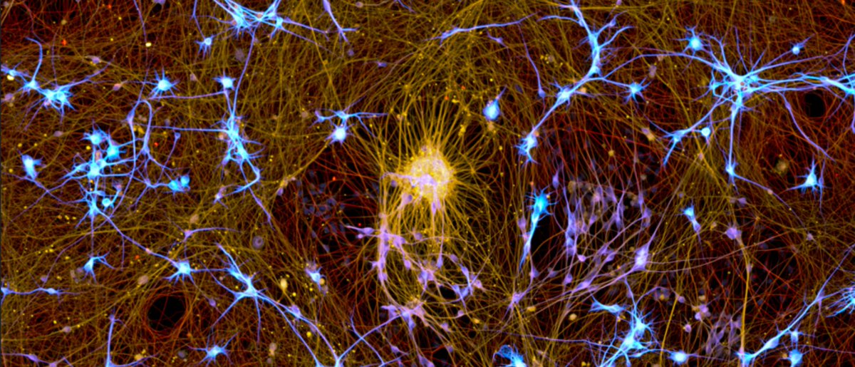
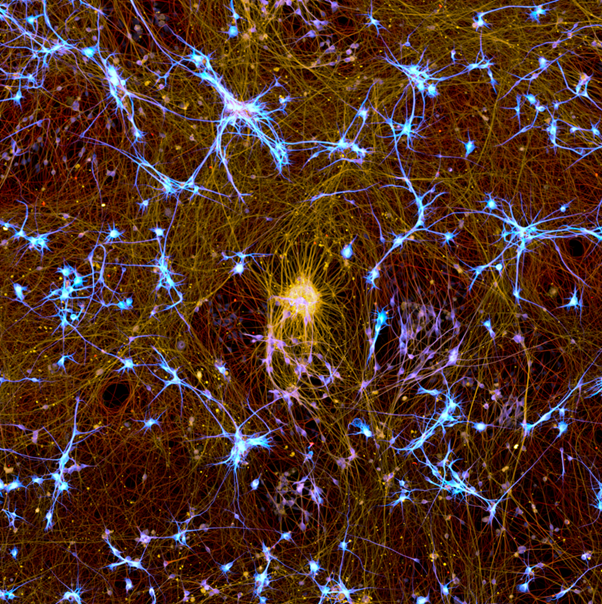
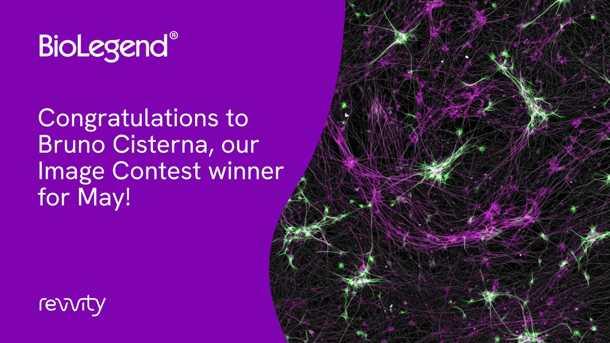
Congrats to Bruno Cisterna (@CisternaBA, Augusta University), our May winner, for their image of Tubulin β 3, Tubulin, and MAP2 staining in human induced pluripotent stem cells. Our contest ends June 30th, so submit your best images soon! https://t.co/dWx7mjwIqn
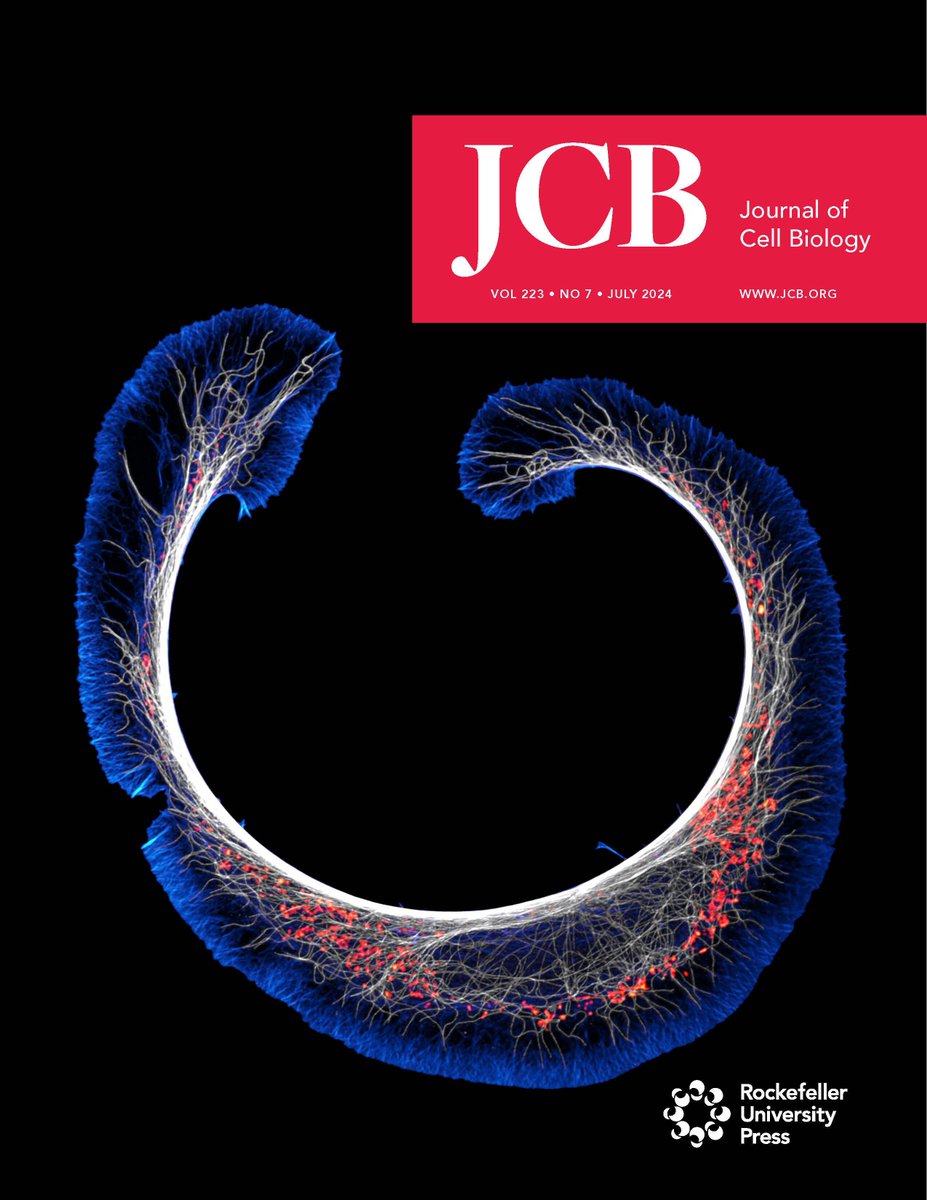
📢 Our 2024 Cover of The Year 📸 goes to this beautiful, super-resolution spinning disk confocal image of a cath.a-differentiated (CAD) cell stained for F-actin, microtubules, & mitochondria. Congrats to @CisternaBA @chillinwithpfn1 and colleagues 🎉 https://t.co/lKKsdgZIPW
Gracias Biología UC por esta excelente nota sobre la historia detrás de nuestra imagen ganadora de la 50ª edición de #NikonSmallWorld 🔬
¡Un honor llevar el nombre del alma mater de mi doctorado a un escenario global!
https://t.co/AB9sjKnH7O
Thank you @thermofisher for choosing my image in the Microscopy Imaging Contest 2024! It’s an honor to share this space with talented scientists https://t.co/Hj0IrRyLS7
Also, check the Q+A session https://t.co/ogDo2HY3K5
More from the Vitriol Lab @chillinwithpfn1
at @MCG_AUG
New #MastersofMicroscopy features Dr. Bruno Cisterna – winner of this year’s #NikonSmallWorld photomicrography contest!
Click to learn about Dr. Cisterna, his long-time passion for #Microscopy, work in the lab of @chillinwithpfn1, and more: https://t.co/F3OCopRTZC
Honored to be among the winners of the 50th anniversary of #NikonSmallWorld competition! 🤩 It's a privilege to showcase the beauty and complexity of the microscopic world. Thank you @NikonSmallWorld! 🔬
https://t.co/ksWUgiDQy8
Our July issue is out: https://t.co/pShVzmEMdf
Cover shows super-resolution spinning disk confocal image of a cath.a-differentiated (CAD) cell stained for F-#actin (blue), #microtubules (white) & #mitochondria (red). By @CisternaBA @chillinwithpfn1 et al. https://t.co/tNDAm0gwbf
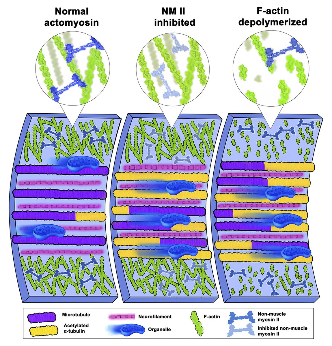
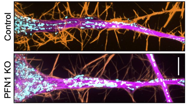
.@CisternaBA, @chillinwithpfn1 @MCG_AUG et al discover that #microtubule number & stability increase following prolonged depletion of profilin 1 or F-#actin due to loss of actomyosin contractility. It is reversible if actomyosin is restored. https://t.co/MwRqnrsHml
#cytoskeleton
Hot from the #preprint!
Former colleague @CisternaBA now @ Vitriol's lab @chillinwithpfn1) show in #neurons & CAD cells how profilin 1 (PFN1) regulates actin (orange) & myo, which changes microtubules (magenta) & #mitochondria (cyan) distribution
Truce in the #CytoskeletonWars?