this is especially prevalent in ADHD btw
you can view ADHD as a disorder/difficulty in loop management
the mind keeps opening new loops while struggling to park or close old ones (dopaminergic dysregulation), so the loops continuously draw cognitive & attentional bandwidth away
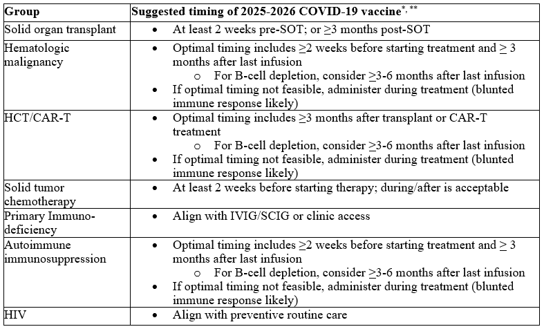
@IDSAInfo 2025 Guidelines on the Use of Vaccines for the Prevention of Seasonal COVID-19, Influenza, and RSV Infections in Immunocompromised Patients
https://t.co/RzIv69FTGb
How to interpret syphilis testing results
Rates of syphilis, including congenital syphilis, have been rising worldwide. Difficult to diagnose given non-specific symptoms and the inability to culture Treponema pallidum on regular media. Various diagnostic methods and complementary testing algorithms have been devised
https://t.co/ygveVPGKkW
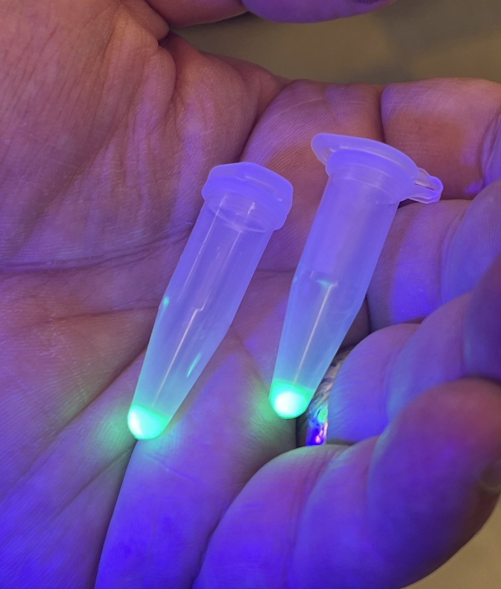
E. coli expressing GFP. Spun down 1 ml of culture, discarded supernatant, and visualized cell pellets with UV light. Thanks to Sebastian S. Cocioba for the strain. @ATinyGreenCell
The bacterial flagellum looks like a simple tail, or whip.
But it’s actually a rotating motor, and perhaps the most sophisticated protein complex nature has ever evolved.
In e. coli, these motors are capable of astonishing speeds; about 15,000 rpm. (The world record, according to one study, is for a Vibrio cell that was “clocked at 100,000 rpm by laser microscopy.)
The flagellum propels the cell forward at speeds of 20-30 microns per second, or roughly 15 body lengths per second. If scaled up to the size of a cheetah, E. coli would *nearly* be the fastest land organism.
The darting movements of a microbe were first observed in 1676 by Antony van Leeuwenhoek, a Dutch cloth merchant. Antony was delighted by the motion of his “animalcules,” writing: “I must say, for my part, that no more pleasant sight has ever yet come before my eye than these many thousands of living creatures, seen all alive in a little drop of water, moving among one another, each several creature having its own proper motion.”
But Leeuwenhoek did not see flagella. He assumed, rather, that these animalcules must be “furnished with paws” instead. Christian Ehrenberg would not properly describe flagella until 1836. But amazingly, all the way up until the 1970s, nobody actually knew how the flagellum spun!
In 1973, there were two competing models people argued over: the helical-wave (bending) model and the rotating (corkscrew) model. The first model suggested that the flagellum whipped back and forth, side-to-side, to propel the cell like paddle. The corkscrew model suggested that the whole flagellum instead spins around like a screw.
In 1974, the corkscrew model finally won out. For two separate studies, scientists affixed flagella to glass slides using antibodies, and watched as the cells spun around and around like corkscrews.
And finally, in just the last year, high-resolution structures of the flagellum have revealed a LOT more about its intricate assembly.
The tail is made from ~20,000 self-assembling copies of a single protein, called flagellin. A “driveshaft,” or rod, spins the tail and is itself made of 26 protein subunits. Each “motor” in E. coli consists of 11 stators, each of which is made from 7 proteins.(Other types of cells have even more stators, and swim with much higher torques.)
The flagellum spins when protons flow into the cell through tiny channels in these stators; akin to water running through a turbine. Each proton makes a small part of the stator change shape and push against the rotor, nudging it forward one step. With dozens of stators working at once, these nudges quickly spin the propeller.
I'm writing an essay for @AsimovPress about this now, and am really enjoying learning about the flagellum and its history. It's an extraordinarily complicated structure, though, and has been a challenge to understand!
A single E. coli cell, placed on a dish, will become 70 billion cells in just 12 hours. That’s exponential growth.
But a new preprint shows that it's possible to engineer E. coli to grow linearly instead, where only one daughter cell continues dividing and the other stops.
First, some context. In nature, there is a bacterium called Mycobacterium smegmatis (initially discovered in 1884 in ulcers scraped from syphilis patients.) M. smegmatis is weird because it divides asymmetrically. These cells grow only from one end, and all their cell wall biosynthesis machinery is located on that one end. So when the cell divides, one daughter gets this machinery and the other gets nothing. The daughter that gets the machinery can keep dividing immediately, but the other daughter has to remake all that machinery from scratch, so its growth is delayed.
E. coli doesn’t grow like this. When it divides, it pinches in the middle and splits everything evenly. Enzymes, metabolites, and proteins get partitioned more or less randomly between the two daughters.
For the new preprint, though, researchers engineered E. coli to behave more like M. smegmatis. Here is how they did it:
First, they deleted a gene called cyaA, which encodes an enzyme (adenylate cyclase) that makes a molecule called cAMP.
cAMP is SUPER IMPORTANT! It is a nutrient sensor that instructs E. coli to switch on genes that help it digest non-glucose carbon sources when glucose is scarce. Without cAMP, E. coli cells growing on alternative carbon sources will starve; they won’t know how to eat the food.
Next, they added back a “split” version of the cyaA gene into the cells. In other words, they split the gene in two so that each half of the enzyme is made separately. Cells can only make cAMP, and thus eat non-glucose carbon sources, if these two halves come together.
To facilitate that “coming together,” the researchers also fused the split cyaA proteins to sticky proteins that clump together, and to a fluorescent protein (to make it easy to track these molecules in the cell.)
So now some interesting things start to happen if you grow E. coli on a growth medium lacking glucose.
As the cell grows, its cyaA “halves” start clumping together into a giant ball. Inside the aggregate, the two enzyme halves come together and make cAMP. And when the cell gets big enough and divides, the clump of cyaA RANDOMLY goes to either daughter cell #1 or #2.
The daughter that gets the aggregate (called PA+ in this paper) can keep dividing. The daughter that doesn’t (PA–) cannot. It still grows a few times — about four divisions — because it inherits some leftover cAMP from its mother. But after that, the metabolite is diluted away, and the cell stops growing.
PA+ cells went through about 23 divisions on average before their aggregate decayed. And the population of cells, as a whole, grew linearly.
This paper is cool because there are many applications where exponential growth is too unpredictable and, perhaps, unsafe. If you want to engineer bacteria to deliver drugs, clean up waste, or live in the gut, you don’t want them to double uncontrollably. This paper shows you can make them expand in a controlled, linear way.
Alas, mutations could break this whole engineered system. A mutation that restores cyaA, for example, would give cells a new way to make cAMP. Mutations that make the aggregates split between daughters would break the asymmetry, too. But still, I really enjoy proof-of-concept engineering papers like this.
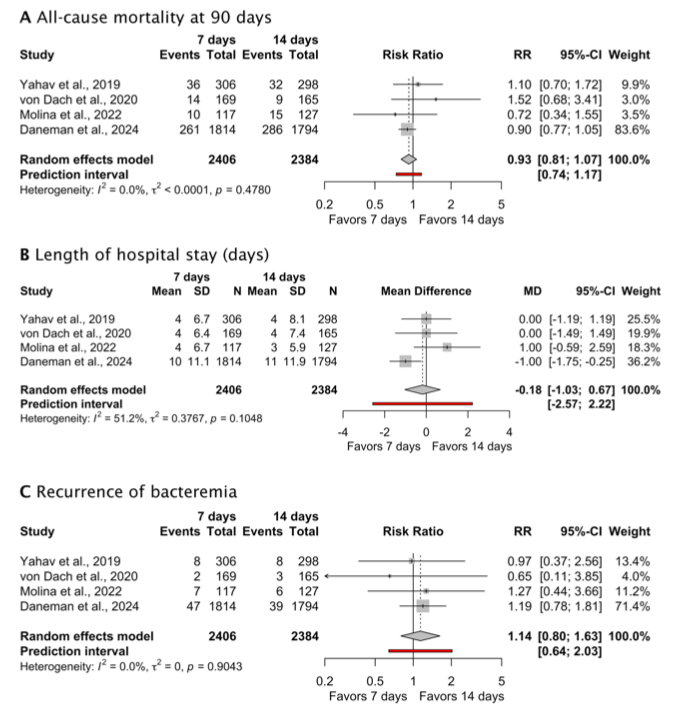
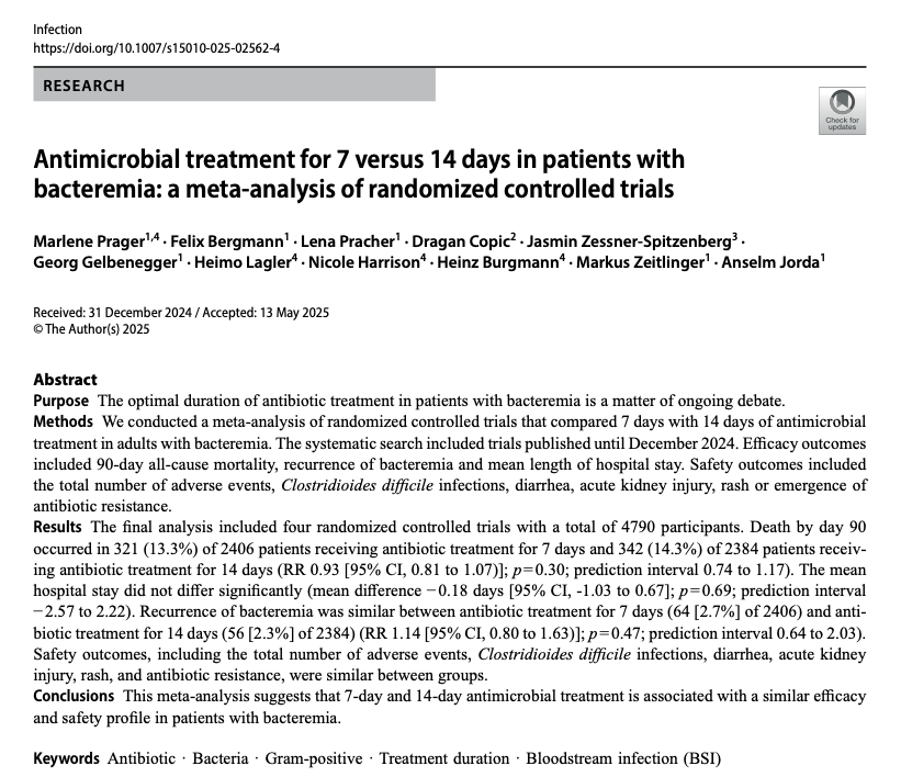
Antimicrobial treatment for 7 versus 14 days in patients with bacteremia: a meta-analysis of randomized controlled trials
🔺Do we need more of these??
https://t.co/3tm7p7RSAn
Updated #IDSAGuidelines! IDSA has issued two new guidelines on abatacept and infliximab for COVID-19, offering evidence-based recommendations for their use in hospitalized patients with moderate to severe disease.
View guidelines: https://t.co/H4KGct57fZ
Resistance mechanism mnemonics!
-Linezolid Resistant Enterococci (bear with me)
The image is 23 cops (police) preventing it from acting
23 - the most common mechanism is 23s rRNA mutation
COP - the other mechanisms are transferable genes. C = cfr +cfr(b), O = optrA, P = poxtA