@AntelmPujol Banda, solo seamos mejores, seamos objetivos, estudiemos más, la IA no se va a parar delante de un paciente tan pronto, no se va a poner delante de un papá asustado, delante de alguien que necesita respuestas que no comprende, solo seamos mejores
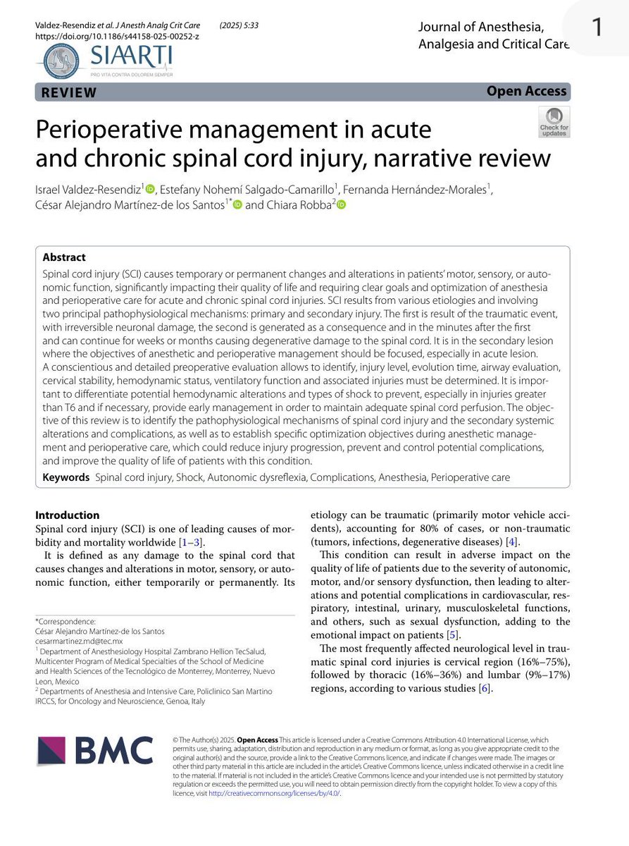
🧠 The first minutes after a spinal cord injury determine much more than neurological recovery. They determine whether secondary injury will amplify or limit the damage.
Primary spinal cord injury is immediate and largely irreversible. Mechanical trauma destroys neurons, blood vessels, and axons within seconds. What follows, however, is a prolonged cascade of ischemia, inflammation, edema, oxidative stress, excitotoxicity, apoptosis, and microvascular dysfunction that can continue for weeks or even months. This secondary injury is where intensive care, anesthesia, and perioperative medicine can make the greatest difference.
This narrative review reminds us that perioperative management should not focus solely on the surgical procedure but on preserving spinal cord physiology. The first priorities are identifying the neurological level of injury, assessing airway stability, recognizing associated trauma, and maintaining adequate spinal cord perfusion. Patients with injuries above T6 deserve particular attention because they are at high risk of neurogenic shock, characterized by hypotension and bradycardia from sympathetic denervation, which can critically reduce spinal cord blood flow if not promptly treated. Importantly, neurogenic shock must be distinguished from spinal shock, a neurological syndrome of transient areflexia and paralysis rather than a circulatory shock state.
Respiratory management is equally crucial. The level of injury predicts respiratory muscle dysfunction, secretion clearance, cough effectiveness, and the need for ventilatory support. Cervical injuries frequently require prolonged ventilation, whereas lower thoracic lesions preserve much of respiratory mechanics. Preventing atelectasis, pneumonia, hypoxemia, and hypercapnia is essential because secondary hypoxia directly worsens spinal cord ischemia.
The review also highlights several practical perioperative principles. Airway management should minimize cervical movement, with video laryngoscopy or fiberoptic intubation performed by experienced operators. Succinylcholine should be avoided after the first 48 hours because of the risk of severe hyperkalemia. Hemodynamic management should prioritize spinal cord perfusion by avoiding hypotension, maintaining normovolemia, and using early vasopressors when necessary, with norepinephrine generally preferred. Current recommendations suggest maintaining a mean arterial pressure between approximately 75-80 mmHg as the lower limit and avoiding active increases above 90-95 mmHg during the first 3 to 7 days after acute injury.
Reference 📚
Valdez-Resendiz I, Salgado-Camarillo EN, Hernández-Morales F, Martínez-de los Santos CA, Robba C. (2025). Perioperative management in acute and chronic spinal cord injury, narrative review. Journal of Anesthesia, Analgesia and Critical Care, 5, 33. https://t.co/zFxOc2pSSq
🫀Twenty-five years of septic shock research have taught us one essential lesson: physiology is more important than protocols.
For years, septic shock resuscitation revolved around correcting isolated variables. We targeted central venous oxygen saturation, serum lactate, mean arterial pressure, or fluid volume as if each represented the entire circulatory system. Landmark trials demonstrated that although these variables provide valuable physiological information, none should be interpreted in isolation. Septic shock is a heterogeneous syndrome, and identical hemodynamic values may reflect completely different pathophysiological states.
This outstanding perspective by Hernandez and colleagues reviews twenty-five years of evidence, from Early Goal Directed Therapy to the recently published ANDROMEDA SHOCK 2 trial. The evolution is striking. Resuscitation has progressively shifted from protocol driven algorithms toward individualized physiological assessment incorporating peripheral perfusion, fluid responsiveness, critical care echocardiography, hemodynamic phenotyping, and repeated bedside reassessment.
Perhaps the most important concept is that every intervention should become a reversible physiological test. Fluids should only be administered if the patient is fluid responsive and likely to tolerate them. Vasopressors should not simply normalize mean arterial pressure, but determine whether increasing perfusion pressure actually improves tissue perfusion. Dobutamine should be guided by cardiac phenotype rather than routine use. Most importantly, each intervention should be followed by immediate reassessment to determine whether it achieved its intended physiological objective.
Capillary refill time emerges as more than a simple bedside sign. Its rapid response to changes in perfusion makes it an attractive marker to guide sequential resuscitation while potentially reducing over-resuscitation. At the same time, lactate, ScvO₂, mean arterial pressure, and fluid balance retain their value when interpreted within the patient's overall physiological context rather than as isolated therapeutic goals.
The authors also remind us that many of the greatest advances in septic shock management have not resulted from new drugs or expensive technologies. Instead, they have come from a deeper understanding of cardiovascular physiology and better application of bedside monitoring. The future is unlikely to be defined by another universal algorithm. It will be defined by personalized, physiology-guided resuscitation tailored to each patient's evolving hemodynamic phenotype.
Reference 📚
Hernández G, Hunsicker O, de Backer D, et al. (2026). Twenty-five years of septic shock hemodynamic resuscitation trials: a conceptual perspective. Critical Care, 30, 400. https://t.co/57pYcSZ5de
"Memoria selectiva para recordar lo bueno, prudencia lógica para no arruinar el presente y optimismo desafiante para encarar el futuro".
Isabel Allende
📷Elliott Erwitt
A new review summarizes platelet-activating anti–platelet factor 4 disorders, including heparin-induced thrombocytopenia and related syndromes, highlighting diagnosis, antibody mechanisms, and treatment strategies. 👉 https://t.co/5mnvrPal1Z
The pathogenesis of vaccine-induced immune thrombocytopenia and thrombosis (VITT) is summarized in Figure 3 (see here). The conventional view — similar to that regarding heparin-induced thrombocytopenia (HIT) — is that a polyanionic vaccine constituent interacts with platelet factor 4 (PF4) to create a neoantigen that results in VITT. However, such a mechanism would imply an iceberg model and also fails to explain why, when VITT occurs, it nearly always does so after the first vaccine dose. The current model is that VITT represents a misdirected recall immune response against the adenovirus core antigen protein VII (pVII) that is potentially enhanced by free DNA within the vaccine. During the strong boosted immune response against pVII, one or more B cells shift antibody specificity from adenovirus pVII to PF4. For this to occur, several factors must converge: first, the B cell produces an antibody that recognizes a distinct epitope on pVII (but which shows structural mimicry with an epitope on PF4); second, the IgG hypervariable-region light-chain haplotype IGLV3-21*02 or *03 is involved; third, the heavy-chain CDR3 region shows a characteristic cluster of negatively charged amino acids (the ED motif); and fourth, a critical somatic hypermutation occurs (K31E), which results in an exchange of a germline positively charged amino acid, lysine (K), to a negatively charged amino acid, glutamic acid (E), at position 31 of the IGLV3-21*02 or *03 allele.
Learn more about VITT and other anti–platelet factor 4 disorders in the Review Article “Platelet-Activating Anti–Platelet Factor 4 Disorders” by Theodore E. Warkentin, MD, and Andreas Greinacher, MD: https://t.co/5mnvrPal1Z