A new publication by our UTSW neuropathology faculty members: The efficacy of immunohistochemistry in the diagnosis of molecular genetic alterations in central nervous system gliomas: Next-generation sequencing of 212 mutations in 112 patients.
DOI: 10.5414/NP301381
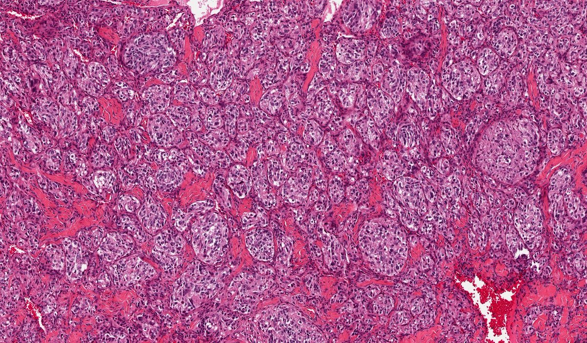
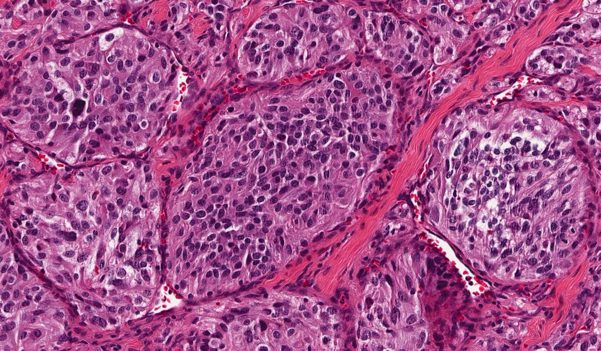
@UTSW_Pathology Paraganglioma! Epithelioid chief cells arranged in tight nests (Zellballen pattern) with salt-and-pepper chromatin, stain with neuroendocrine markers and keratin. Surrounding sustentacular cells stain with S-100. Stains not shown.
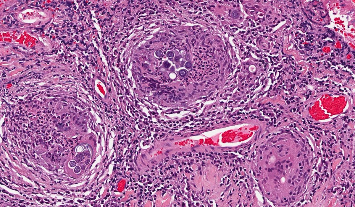
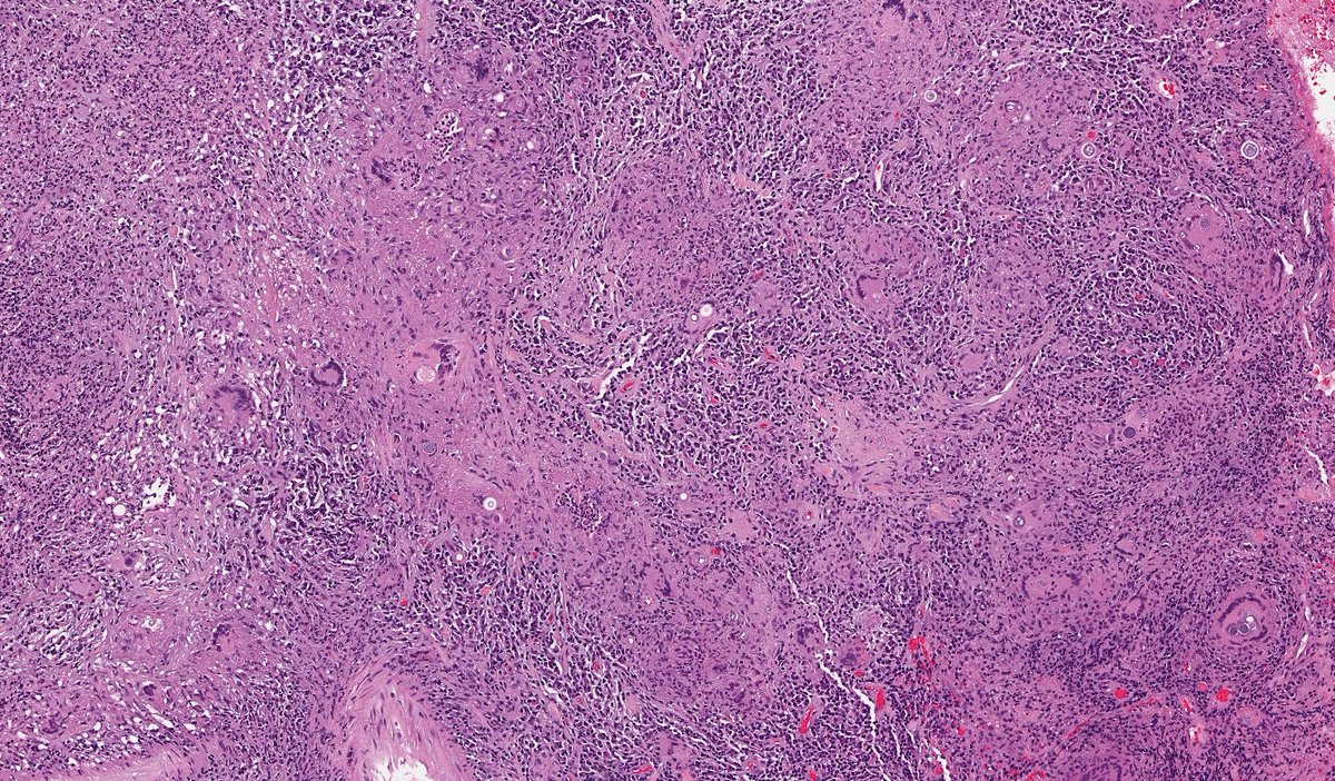
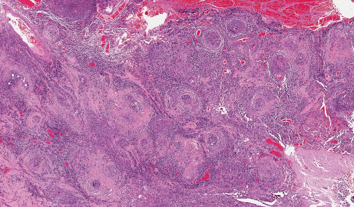
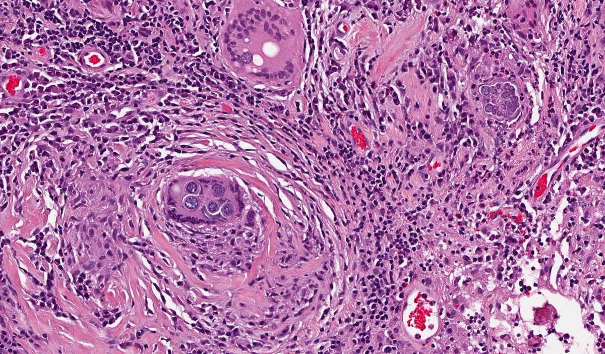
This is Coccidioides which is endemic in southern USA, Central and South America. CNS presentation is basal meningitis, paravertebral abscess, or intracerebral lesions. Large yeast ~40 um diameter containing numerous tiny endospores here. Thanks for the differentials!
@NorganAndrew Granulomatous parenchymal reaction in an immunocompromised patient certainly raises a possibility of Blasto. The fungi are ~8-15 um diameter with a typically broad-based budding, which is not seen in this case. There should also not be endospores like we see in this case.
@SuntreaHammer Strongy most commonly presents as a purulent meningitis (I didn’t explicitly say this is brain parenchyma). When filariform larvae are present, microinfarcts can occur from small vessel occlusion. Granulomatous inflammation can also occur in some cases, so this is a fair guess!
@ricardocedenomd Granulomatous parenchymal reaction in an immunocompromised patient certainly raises a possibility of Blasto. The fungi are ~8-15 um diameter with typically broad-based budding, which is not seen in this case. There should also not be endospores like we see in this case.