Taking #CLEM into the 21st century, read all about it here https://t.co/I3xzyc4pnC watch all the videos here https://t.co/JoLMJ9r1wp and extra points for anyone that even attempts to read the full supplement. Great #science when #biology and #physics meet
Correlative two color 3D super-resolution single molecule localization microscopy and block face electron microscopy of mitochondria and the endoplasmic reticulum in vitreously frozen COS-7 cells https://t.co/yWLSkjoPuZ
(1/x) We are excited to introduce VIPS (Volumetric Imaging of biological specimens via Photochemical Sectioning) in our preprint! VIPS uses a light-based process called “photochemical sectioning” to achieve petabyte-scale high/super-resolution imaging: https://t.co/Ubn7o4ocsa
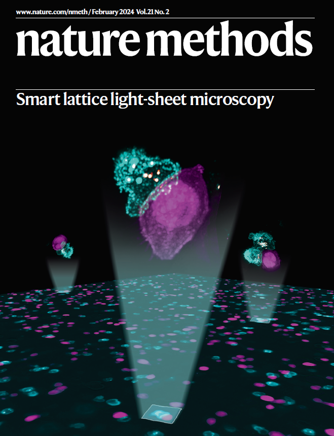
We're very pleased to announce the February issue of Nature Methods, which includes a "mini-Focus" follow-up to our July 2023 Focus Issue on the future of bioimage analysis! (please see thread) https://t.co/cfkIHYOovc
✅💡 very happy to have this paper finally out. Chris and I started working on this project in 2017…so great to get this across the line at last!
https://t.co/zzUMuW11YF
Cellular organelles exchange molecular information through transient contact sites, which are sensitive to experimental analysis. Reporting @nature, scientists use advanced microscopy to map and analyse dynamic subdomains at ER-mitochondria contact sites.
https://t.co/6Fvy4foVGV
Excited to share our comparison of Xenium and CosMx data! I feel like a lot of labs/core facilities have made similar observations internally, so we're really glad to put this out for the broader community. https://t.co/avvKe0QDBT 1/n
Muyuan has been working on this for some time, it's cool to see a manuscript: How to create full, atomic resolution scenes of biomolecules in Unreal Engine and fly around them in real-time. Really cool stuff with untapped potential.
https://t.co/dWQ6nhRnNJ
https://t.co/oRDsvwECgj
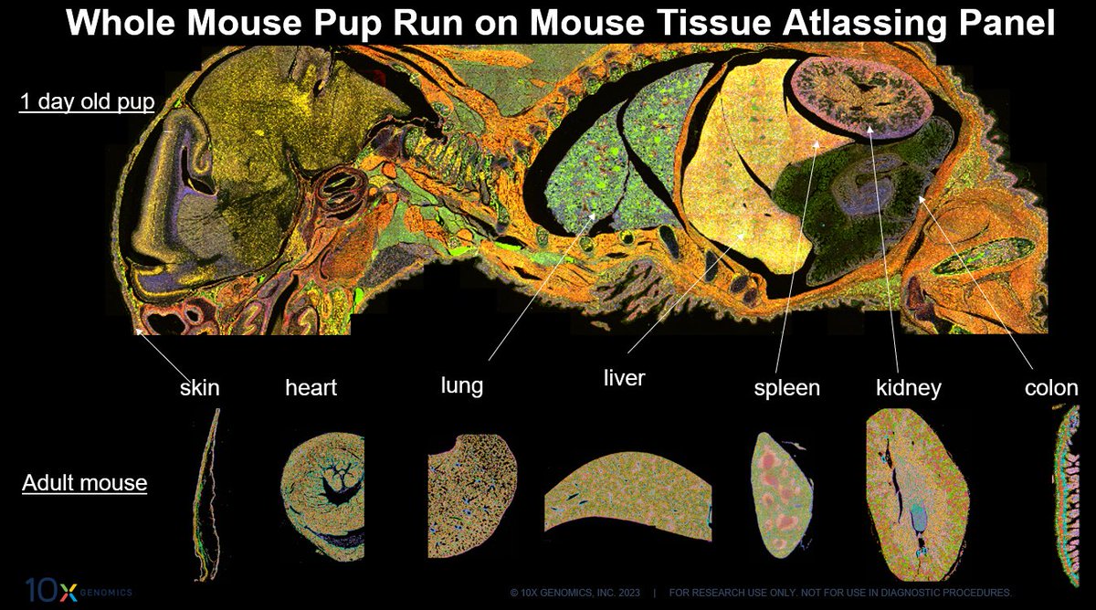
Biology is beautiful—and Xenium proves it by bringing it to life with the new Mouse Tissue Atlasing Panel. Learn how #singlecellspatial imaging can help you uncover the splendor of your sample here: https://t.co/tJ4IBtvlbK
If you are imaging more than one fluorophore in your specimen, be sure to perform the correct controls to ensure your results are not confounded by bleedthrough or autofluorescence!
https://t.co/xHNVeL8Cv1
What if you could explore 1,000s of your genes at subcellular resolution & get deeper cellular and spatial insights? What if you could do it with near-instant data after a run?
Don’t wonder: find out how Xenium can take your ‘what ifs’ to ‘what next’. https://t.co/CfvisUz6nX
Our paper on fluorescence lifetime voltage imaging of neurons is out! We combine kilohertz frame rate, micrometer spatial scale, and picosecond lifetime resolution in vivo. Check out the videos and original thread below. @ChengHuangThu
https://t.co/Ek4OyPkGIs
If you like building cool light-sheet microscopes, this is for you. The benchtop mesoSPIM @MesoSpim is faster, has better optics, larger field of view. Works for whole mouse, human cortex and more.
@voigtvision @FritjofHelmchen @nvladimus
https://t.co/gtelkNf7Pk
The nd2 package (Nikon NIS elements file reader for python) just got a major internal refactor. v0.6.0 reimplements the outputs of the official SDK in pure python. (No more wrapping of c code)
Speed is improved, and it’s easier to install anywhere now.
https://t.co/DE8c6sIZOC