I am very happy to have our latest paper 'Intra- and intersession reproducibility of artificial scotoma pRF mapping results at ultra-high fields' published in #eNeuro@SfNJournals
https://t.co/TlwekEw79v
We further present CON-pRF, a fully containerized analysis pipeline designed to enhance reproducibility and comparability across studies.
Huge thanks to my co-authors Michael Woletz, Pedro M. Paz-Alonso, @cwindfMRI, and @garilerma for their amazing contributions!
🚨 New preprint alert! 🚨
Check out our latest work on retinotopic mapping! We explore biases in pRF mapping and show how upsampling affects pRF coverage in the central visual field.
Read more: https://t.co/hX5FMTmfPn
#fMRI#Neuroimaging#pRF#OpenScience
While widely assumed, few studies actually show a direct link between pRFs & spatial tuning in visual cortex. Here we found that adapting to spatial frequency modulates pRF sizes along with SF perception.
By Ecem Altan w/ @StevenDakin & Catherin Morgan
https://t.co/FheGLGOIl5
@sampendu@willjharrison@neuroMDL We’re indeed developing a GPU-powered Python pRF tool that drastically cuts analysis time from several hours to just minutes. Still under development, my college Siddharth Mittal will present it at VSS and OHBM. Hope to connect there!
🔬 New study on TBI (Traumatic Brain Injury)! We systematically reviewed the effectiveness of non-invasive brain stimulation techniques (TMS & tDCS) on post-TBI symptoms. #AlbertoGalimberti@MartinTik1@GPellegrino_ 🧠 #TBI
https://t.co/jChTynF8JZ
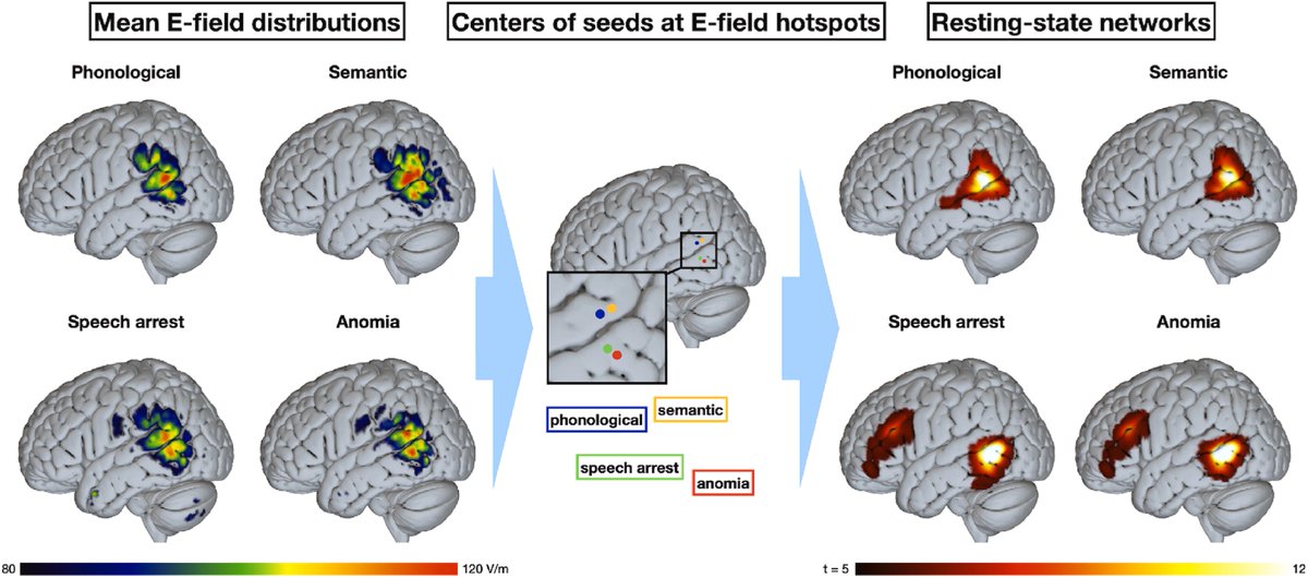
Thrilled to work with talented colleagues like @MarVasileiadi, who used navigated TMS to explore how different subregions of the temporal gyrus affect language production 🧲🧠
👉 Peaks for phonological & semantic errors in STG
👉 Peaks for anomia and speech arrest were in MTG
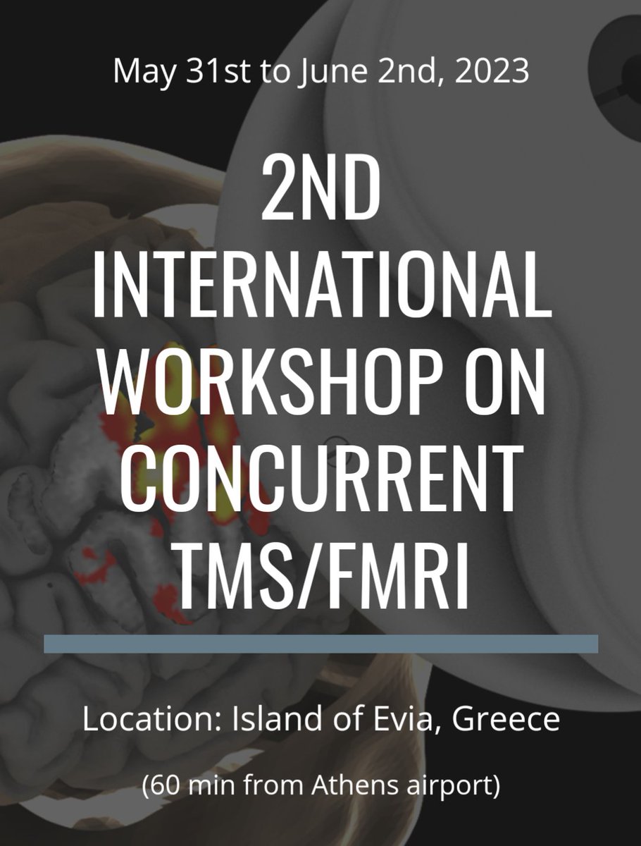
📢 Deadline extended to Apr 24 for the 2nd Intl TMS/fMRI Workshop! Don't miss this opportunity to connect, collaborate, and get inspired by experts in the field. Join us in beautiful Evia, Greece, for an unforgettable experience! 🌊☀️🧠 Register now! 👉 https://t.co/fPR3IBEmXp
Our results are consistent with those of previous studies conducted on healthy subjects (https://t.co/RJL9E4veof).
Thanks to our team and all collaborators!
M. Pawloff, M. Ritter, @cwindfMRI, U. Schmidt-Erfurth, @garilerma
We investigated the impact of different stimulus patterns on pRF mapping results in patients with retinal disease. Our findings demonstrate strong differences in the distribution of pRF centres between bar and wedge/ring stimulation.
Markus Ritter et al. @MedUni_Wien quantitatively assess the performance of different visual stimulation approaches for mapping visual field coverage.
https://t.co/B9MoJaVWOq
We observed a higher pRF density in foveal areas and reduced pRF sizes with wedge/ring stimulation. However, both stimuli are highly effective in detecting visual field defects. pRF mapping even offers superior scotoma detection compared to microperimetry in some cases.
3...2...1...Our #PhDCall is officially open! 📢💥 Are you looking for a #PhD position and want to become an expert in human-centered technologies & sciences? #MedUniVienna got you covered. Scroll through our open #PhDpositions and apply now! ➡️ https://t.co/NBCDLkGEqB
#PhDLife
Excited to be at @bcbl_ in beautiful San Sebastián for my @EMBO Short-Term Fellowship, working with @garilerma on new theories in visual neuroscience and pRF mapping.
I am very happy to have our latest paper 'Intra- and intersession reproducibility of artificial scotoma pRF mapping results at ultra-high fields' published in #eNeuro@SfNJournals
https://t.co/TlwekEw79v
Thus, we have proven that pRF mapping is a highly reproducible brain mapping method that is also very robust to scotoma data. Furthermore, the results provide a foundation for longitudinal pRF mapping studies in patients suffering from partial visual field loss.