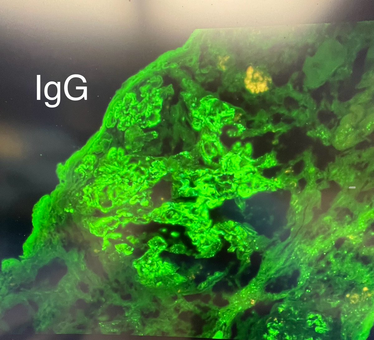
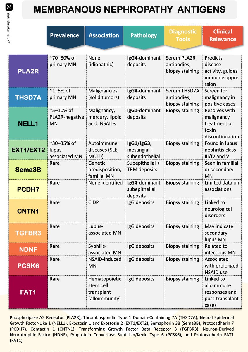
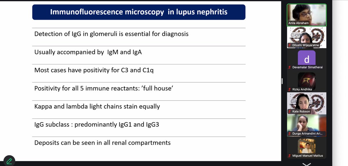
🔔Check out this Chapter 👉 Approach to Glomerular Disease by Dr. Maria Fernanda Zavala- Miranda. and Dr. Juan M. Mejia- Vilet
Access the chapter here: https://t.co/2kgsgZd0q5
#GlomPedia#GlomCon
🧠 Understanding Papillary Thyroid Carcinoma (PTC) Variants 🦋
Dive into the diverse world of PTC subtypes with this detailed breakdown of histopathological features, molecular alterations, and clinical insights. 🩺✨
#ThyroidCancer#PathologyEducation#MedicalLearning
1/6🩺 Clinical Pathology Case @MGHKidneys @mgh_transplant@MGHPathology
Today, we reviewed a 50-year-old man with end-stage kidney disease, who received a kidney transplant in 2020.
🕒 Fast forward 4 years: He presented with rejection, and here’s what we learned! 🧵👇
According to the ISN/RPS 2003 classification of lupus nephritis, membranous lupus nephritis (class V) is characterized by the presence of global or segmental subepithelial immune deposits with or without associated mesangial deposits or mesangial hypercellularity. Membranous lupus nephritis may occur in isolation or may coexist with other forms of active lupus nephritis such as focal (class III) or diffuse (class IV) lupus nephritis. In the presence of active glomerular lesions, scattered subepithelial deposits are common and are not diagnostic of membranous lupus nephritis (class V). In fact, in order to make a diagnosis of membranous lupus nephritis (class V) in the presence of a class III or class IV lupus nephritis, it is required for subepithelial deposits to be present in greater than 50% of the tuft of greater than 50% of the glomeruli by light microscopy or immunofluorescence. Figure 1 shows a glomerulus from a patient with known systemic lupus erythematosus displaying active lesions including endocapillary proliferation and endocapillary hyaline thrombi. Furthermore, electron microscopy (Fig 2) from the same biopsy shows frequent, large subendothelial deposits (red arrows) and few scattered subepithelial deposits (blue arrows). Given the presence of active glomerular lesions and involvement of less than 50% of the tuft by the subepithelial deposits, this case does not meet diagnostic criteria for membranous lupus nephritis (class V).
#TeachingPoints #kidneypath #renal
Patient with acute kidney injury following vancomycin treatment for pyelonephritis. Numerous unusual casts (no LC restriction, myo/hemoglobin and chromo negative). EM shows mixture of uromodulin protein and precipitate. C/W vancomycin cast nephropathy.
#renalpath#nephrology
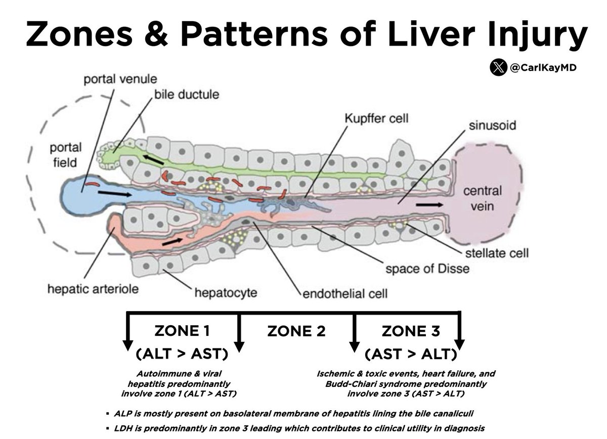
Zones & Patterns of Liver Injury
Zone 1 (ALT > AST)
▪️Autoimmune hepatitis
▪️Viral hepatitis
Zone 3 (AST > ALT + ⬆️ LDH)
▪️Ischemia
▪️Toxic events
▪️Heart failure
▪️Budd-Chiari syndrome
Note: ALP is predominant in basolateral membrane
#LiverTwitter 👊
Breast-Cystic Neutrophilic Granulomatous Mastitis (CNGM)
Histologic criteria
1-Lobulocentric inflammation
2-Cystic spaces rimmed by neutrophils
3-Non-necrotizing granulomas
4-Gram stain with gram positive corynebacteria
If you have at least 2 of the first 3 criteria and a positive Gram stain, you can sign it as "Findings consistent with CNGM" and add a comment like part A of the 1st pic. If the Gram stain is negative for bacteria but you have 2 of the 3 histologic criteria, you can sign it as "Findings suggestive of CNGM" and add a comment like part B of the 1st pic.
Dr Turashvili CCSP24 #pathology #pathologists #PathTwitter #PathX
1.
Introducing a new thread: super basic lung pathology for PGY1s, with arrows. If you are a PGY1, leave a comment and I will tag you in a future post.
A hamartoma is a disorganized mix of overgrown normal tissues (in their normal location) that creates a mass. Good news: they are benign!
#pathology #pulmpath #tweetorial #pathtweetorial #sblppgy1
This is difficult & rare.
Suspicion is key.
LM-large glomeruli, appear hypercellular, but a closer look & you see foam cells.
Stain for CD68 & glomerular capillaries light up & are filled with CD68+ macrophages.
IF/EM essentially negative.
Dx: Histiocytic glomerulopathy
1/3
The Nephrology Board Review & Certification Exams Edition (Part 1)- high yield content for exams not only in the US but also worldwide (in addition to being pertinent clinically). Contents below @akdhjournal@NKF_NephPros@CharuThakar@jwilliamMD@svuh
https://t.co/4wXshGo2eR
The ISN/RPS 2004 classification of lupus nephritis is entirely based on glomerular changes. Depending on the presence or absence of active and/or chronic glomerular lesions, cases are allocated into one (and occasionally two) of six possible classes. The most commonly encountered active glomerular lesions in clinical practice are in the form of endocapillary or extracapillary proliferation, fibrinoid necrosis or cellular crescents. However, deposition of large subendothelial immune deposits, even in the absence additional proliferative/necrotizing lesions is sufficient to classify a case as class III or IV, depending on the number of involved glomeruli (Fig 1-3). These deposits, termed “wire loops”, appear as glassy eosinophilic, strongly PAS-positive, fuchsinophilic and non-argyrophilic material, which involves the circumference of a capillary loop (Fig 1 & Fig 3). When these same deposits extend into the lumen of a loop, and are sectioned en face, they appear as endocapillary thrombi and are thus called “hyaline thrombi” (Fig 2). The nature of these deposits may be further confirmed by immunofluorescence and/or electron microscopy.
#TeachingPoints #kidneypath #renal
@arkanalabs Focal segmental necrotizing GN with neutrophilic tubulitis, possibly ANCA associated, infection related, IgA related or autoimmune mediated GN.
Immunotactoid glomerulopathy (ITG): 45-yr old with hematuria, proteinuria, Non-Hodgkin lymphoma
LM: MPGN pattern
IF: IgG kappa
Subtype: IgG1
EM: microtubular deposits along capillary wall
IgG deposits are usually monoclonal (monoclonal ITG) & associated with a lymphoma. 1/2