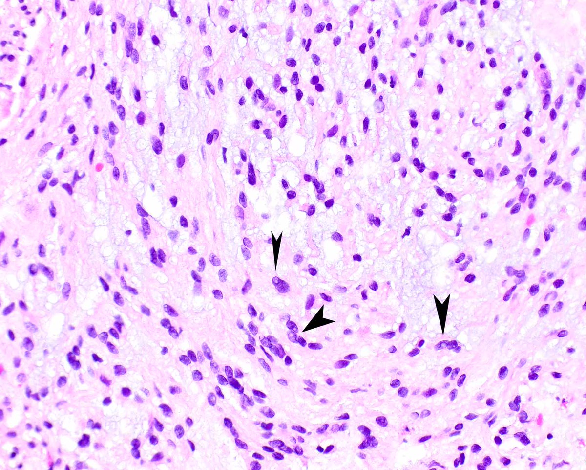
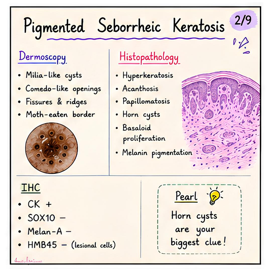
A colonic epithelioid schwannoma (these lesions have been reported using several terms). Note the intranuclear cytoplasmic invaginations (arrows).
Lewin MR, et al. Mucosal benign epithelioid nerve sheath tumors. Am J Surg Pathol. 2005 Oct;29(10):1310-5. PMID: 16160473.
🌍 On #WorldCancerDay, revisit this study on DICER1 mutations in Bethesda category II, III, and IV thyroid nodules: A mutually exclusive relationship with BRAFV600E mutation
https://t.co/l5wiC5rVd4
#CytoPath
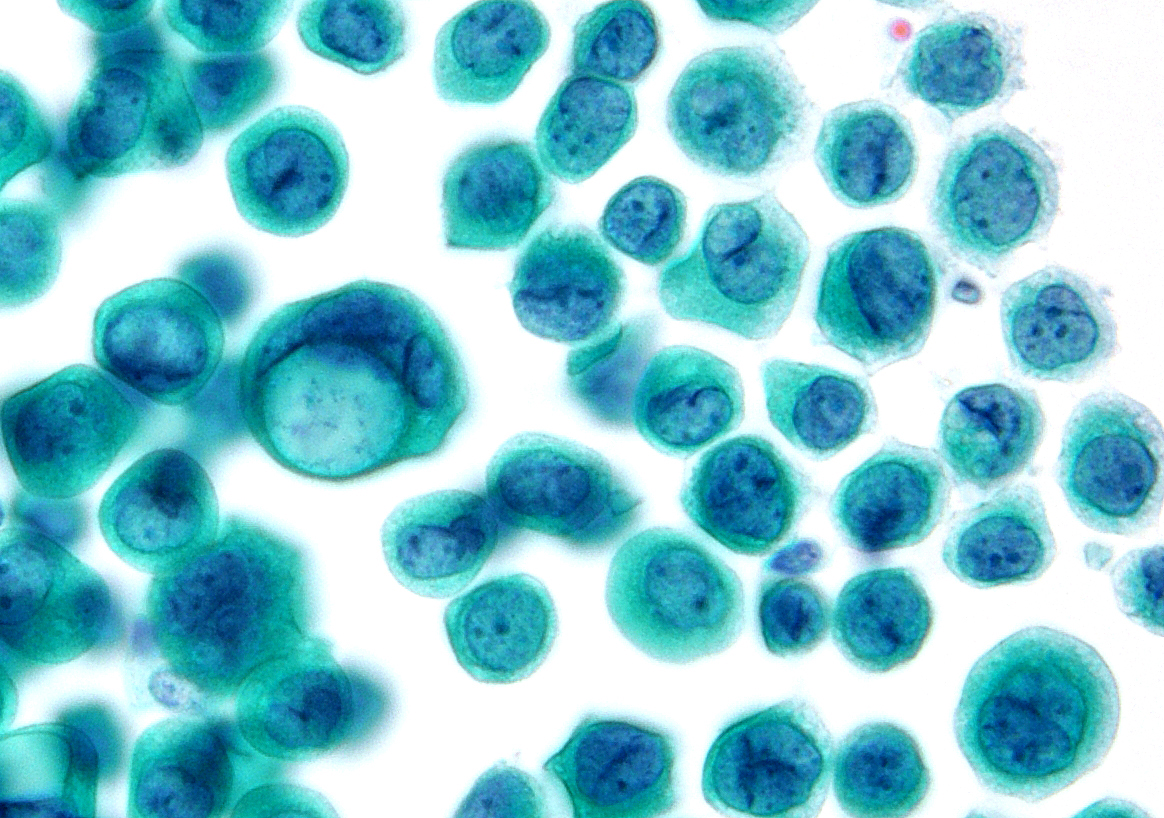
"A pure culture of malignant cells in this ascitic fluid" - Primary site is NOT gastric or lobular breast. The patient had a known history of a "high-grade urothelial carcinoma with focal glandular differentiation and prominent signet-ring cell morphology".
🔬 Prostatic Ductal Adenocarcinoma: Is There a Clear Cell Variant?
📍 Morphological review focusing on rare patterns
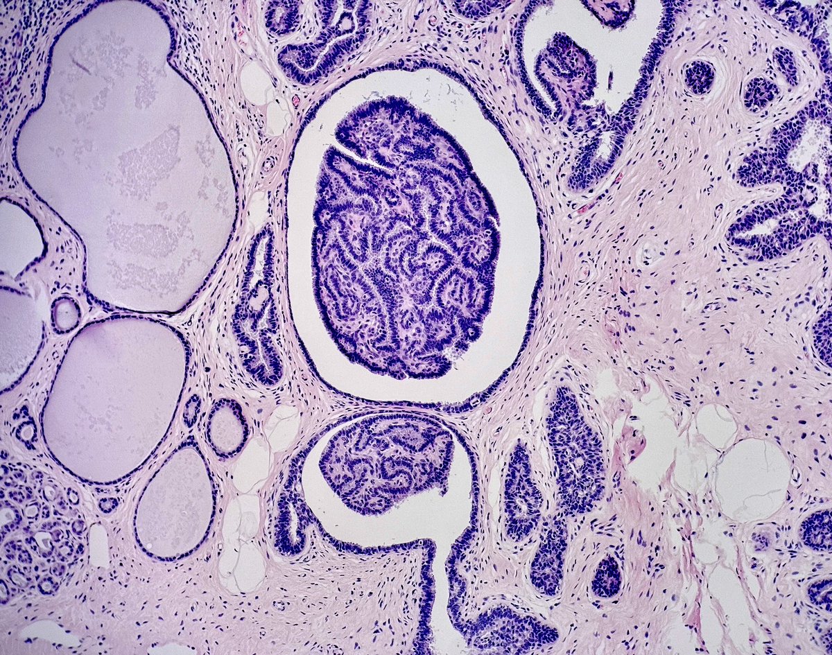
Prostatic ductal adenocarcinoma is a rare and aggressive form of prostate cancer, accounting for only 0.2–0.4% of pure cases. It is classically characterized by papillary and cribriform architecture, lined by tall, pseudostratified columnar epithelium with elongated nuclei and prominent nucleoli.
📌 But could a clear cell variant exist?
Although not formally recognized in current classifications (WHO 2022), there are morphological clues and case reports suggesting this possibility, particularly in the context of unusual differentiation.
🧠 Main morphological patterns
Classic: papillary and cribriform with amphophilic or pale cytoplasm.
PIN-like: flat or tufted architecture resembling high-grade prostatic intraepithelial neoplasia (PIN).
🔍 Rare patterns described (Lee et al., Pathology, 2010):
Foamy gland ductal: papillary/cribriform architecture with foamy, pale cytoplasm (mimicking clear cells).
Mucinous: intracellular and extracellular mucin production.
Micropapillary: slender epithelial projections lacking fibrovascular cores.
Cystic papillary: cystic areas lined by papillary structures.
Paneth cell-like: eosinophilic neuroendocrine granules in columnar cells.
🧬 Prostatic origin was confirmed in these rare cases via positive IHC for PSA, PSMA, and P501S, with negative basal cell markers (p63, HMWCK).
📉 Although some of these variants followed an indolent course, the ductal component is generally associated with higher aggressiveness, including unusual visceral metastases (e.g., liver, lung, penis, testis) and reduced responsiveness to hormonal therapy.
❓What about clear cells?
Even though a “clear cell variant” is not officially recognized, the clear or foamy cytoplasm observed in some ductal adenocarcinomas (notably the “foamy gland” type) raises the possibility that this cytoplasmic pattern may represent a distinct subtype or degenerative phenomenon.
Such cases require careful differential diagnosis from urothelial, renal, or colorectal carcinomas with similar features.
👨⚕️ Conclusion:
There is room to explore the possibility of clear cell morphology in prostatic ductal adenocarcinoma, especially in light of the rare patterns already described. This discussion is important not only for differential diagnosis but also to expand our understanding of the morphological and molecular plasticity of prostatic tumors.
📚 Key references:
Lee et al. Pathology. 2010;42(4):319–324.
WHO Classification of Tumours of the Urinary System and Male Genital Organs, 5th Ed. (2022).
#MedicalEducation #NotasDePatologia
⚠️ Disclaimer: This content is intended for educational purposes only and should not be considered a substitute for professional medical advice, diagnosis, or treatment.
Pyloric gland adenoma in uninflamed gastric body. There is an association with autoimmune gastritis (pyloric metaplasia progressing to pyloric adenoma) but this patient does not have it. Note the appearance of the surface of the adenoma versus the adjoining foveolar epithelium.
Membranous nephropathy has undergone a paradigm shift due to the discovery of unique MN antigens.
MN has gone from idiopathic to ➡️ primary vs. secondary ➡️ where an antigen can be detected in ~80% of MN.
This is a comprehensive review of each antigen.
https://t.co/RJFApwsvG0
Differential diagnosis of carcinoma with squamous differentiation in the Thyroid Gland
-Not every squamous cell carcinoma in the thyroid is anaplastic carcinoma
Dr. Wiswanathan #USCAP25#PathX#pathology
Olfactory neuroblastoma:
- Nests of small round blue cells with salt and pepper chromatin.
- May have Homer-Wright rosettes, or arrangements of tumor cells around neuropil (blue box).
See more annotated features at: https://t.co/MFdtMMK9GH
MONCKEBERG’S ARTERIOSCLEROSIS- dystrophic calcification in the tunica media of medium sized muscular arteries.
BMP-2 and RUNX2 are the osteogenic markers expressed in vascular smooth muscle cells.
🟣 Merkel Cell Carcinoma (MCC): A Rare but Aggressive Skin Cancer 🟣
📍 Also known as primary cutaneous neuroendocrine carcinoma.
🔬 Definition
Merkel cell carcinoma (MCC) is a rare and aggressive primary cutaneous neuroendocrine carcinoma, often presenting as a fast-growing violaceous or flesh-colored nodule in sun-exposed skin.
📈 Epidemiology
Predominantly affects elderly, fair-skinned individuals (median age: 75).
Incidence:
• Europe: 0.13 per 100,000
• USA: 0.79 per 100,000
• Australia: 1.6 per 100,000
Increased risk in immunosuppressed patients (e.g., HIV, organ transplant, CLL).
🦠 Etiology & Pathogenesis
Two major pathways:
1️⃣ MCPyV-positive MCC: Clonal integration of Merkel cell polyomavirus
2️⃣ MCPyV-negative MCC: Strongly associated with UV-induced DNA damage, often with TP53 and RB1 mutations and high mutational burden.
MCPyV-negative tumours tend to be more aggressive.
🧪 Histopathology
Dermal “blue cell” tumour with neuroendocrine cytology:
• Fine “salt and pepper” chromatin
• High mitotic index and apoptosis
• Frequent lymphovascular invasion
May rarely appear intraepidermally (epidermotropic) or as MCC in situ.
🧬 Immunohistochemistry
Positive: CK20 (perinuclear dot-like), synaptophysin, chromogranin, CD56, INSM1, neurofilament, SATB2
Negative: TTF-1 (helps exclude pulmonary origin)
MCPyV status assessed by immunostaining (e.g., CM2B4 antibody)
⚠️ Differential diagnosis
Includes:
Basal cell carcinoma
Small cell melanoma
Cutaneous lymphoma
Metastatic neuroendocrine carcinoma
Cutaneous Ewing sarcoma
Atypical fibroxanthoma
📊 Staging (AJCC/UICC TNM8)
Stage I: Tumour < 2 cm
Stage II: Tumour > 2 cm
Stage III: Regional lymph node involvement
Stage IV: Distant metastasis
Pathologic staging includes size and invasion into deep structures.
📉 Prognosis
5-year overall survival:
• Localized: 51%
• Regional nodes: 35%
• Distant metastasis: 14%
MCPyV-positive tumours have better prognosis.
Interestingly, patients presenting with nodal metastasis without skin lesion may fare better than those with known cutaneous primary.
🧬 Treatment & Advances
MCC is radiosensitive: radiation plays a key role in local control.
Immunotherapy (e.g., anti-PD-1/PD-L1) has improved outcomes in advanced disease.
📚 Key references:
WHO Classification of Skin Tumours, 4th ed., 2018
Harms PW et al., Nat Rev Clin Oncol, 2018 (PMID: 27592804)
Paulson KG et al., J Natl Cancer Inst, 2018 (PMID: 28423400)
Nghiem P et al., NEJM, 2016 (PMID: 27592804)
Paulson KG et al., J Clin Oncol, 2011 (PMID: 21865945)
Goh G et al., Cell, 2016 (PMID: 26022453)
📚 #MerkelCellCarcinoma #NeuroendocrineSkinTumour #DermPath #Oncology #MedicalEducation #NotasDePatologia
⚠️ Disclaimer: This content is intended for educational purposes only and should not be considered a substitute for professional medical advice, diagnosis, or treatment.
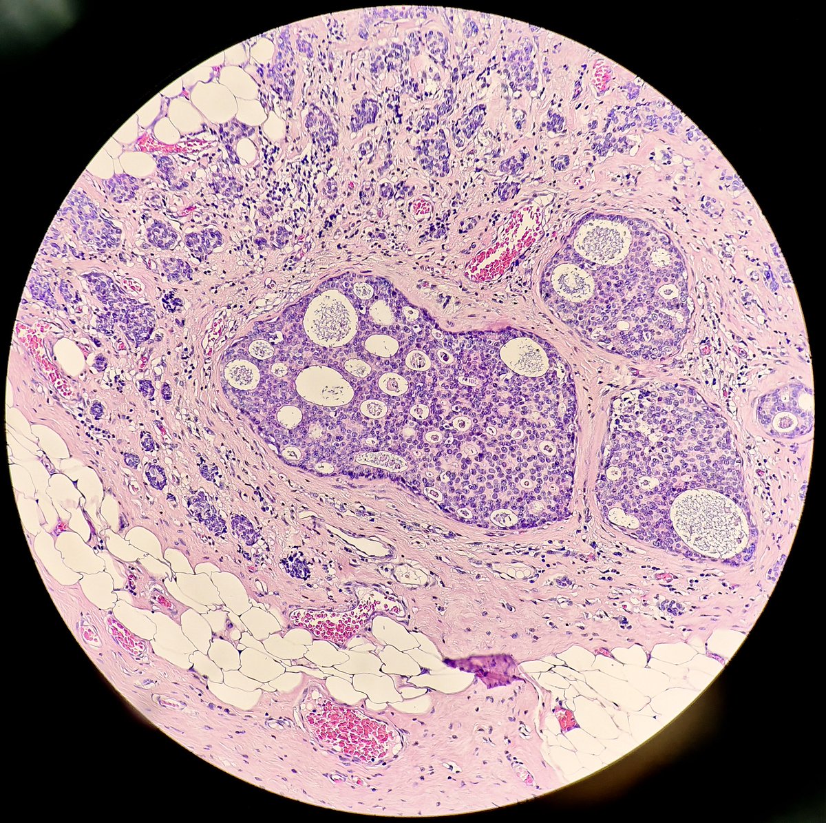
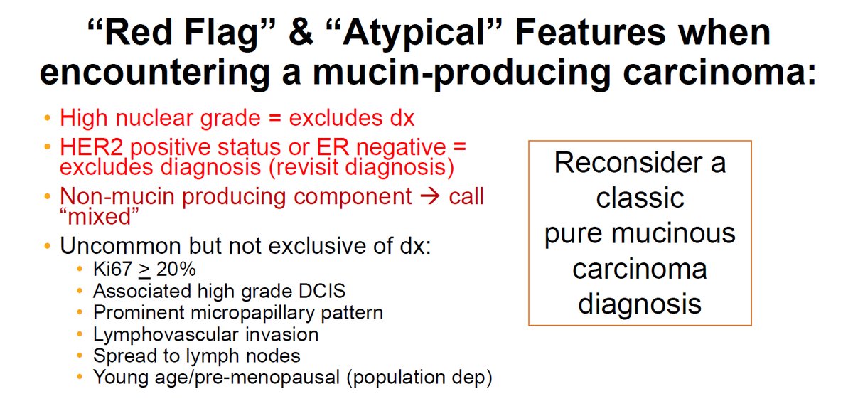
Reconsider a classic pure Mucinous Carcinoma diagnosis if you see red flag/atypical features (pic 1)
-It should not be High grade
-It should be pure (classified on excision specimen)
-It should be HER2 negative and ER positive
Breast Evening Specialty Conference-Dr. Allison #USCAP25 #PathX #pathology