Quiz: Linear radiopaque material tracking along both sides of the skull on CT. What is it?
Answer: Post-procedural changes from middle meningeal artery embolization for chronic subdural hematoma.
Why we do it
•Chronic subdural hematoma is not just “old blood.”
•It is a biologically active membrane disease.
•Outer neomembrane develops along the dura.
•It is vascular, fragile, and fed largely by branches of the middle meningeal artery.
•Recurrent microbleeds and exudation maintain or enlarge the collection.
•Embolization shuts this supply → less rebleeding → lower recurrence.
Where it fits clinically
•Adjunct to burr-hole evacuation to reduce recurrence
•Standalone in selected patients (mild symptoms, high surgical risk)
•Recurrent or bilateral chronic SDH
•Increasing use with growing evidence, but still evolving practice patterns
—Pearls, pitfalls and wisdom from my reporting list
This elderly woman presented with difficulty in speaking, which had progressively worsened over a period of 4 days.
Diagnosis?
1. Sublingual epidermoid cyst
2. Carcinoma of the tongue
3. Beckwith-Wiedemann syndrome
4. Diphtheria
5. Tuberculosis of the tongue
Posterior pituitary bright spot.
It is a T1 finding.
Often not seen on FLAIR.
So absence on FLAIR does not mean true absence.
Pearl: Judge it on T1.
Pitfall: Calling it absent on FLAIR.
—Today’s reporting list
79/M. Left otitis media with left facial palsy and hearing loss.
In suspected malignant otitis externa (skull base osteomyelitis), the earliest clue is often not bone. It is loss of normal fat.
Subtle asymmetry.
Blurring of intermuscular planes.
Fat disappearing where it should be crisp.
Around the stylomastoid foramen.Within the masticator space. Along the skull base.
Easy to overlook.
Easy to dismiss.
By the time bone is destroyed, you are already late.
Reporting pearl:
Loss of deep skull base fat planes should be considered skull base osteomyelitis until proven otherwise, even if the bone looks intact.
—Pearls, pitfalls and wisdom from today’s reporting list
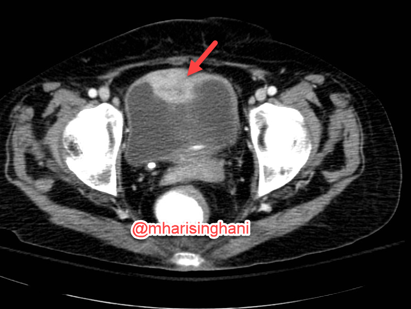
No testicular flow on ultrasound, but the scrotum isn't tender. What's the diagnosis? Watch Shweta Bhatt, MD break it down in this session from Mastering Genitourinary Imaging & Interventions.
Register now for access to past and upcoming sessions: https://t.co/dQh9JsyjzP
🌍Live from IPNTN Season VII, Session 1🌍
Head and Neck Pathologies, Take 5! with @amyfjuliano
💡Think carefully about the wording of reports! Mastoiditis is a clinical diagnosis, coalescent mastoiditis and other complications of mastoiditis should be assessed by imaging. And remember, not all mastoid opacification is infectious.