Rare case...2 Level Corpectomy for treatment of a ruptured Anterior Radiculomedullary Artery aneurysm, good example of looking for alternative causes of SAH when the initial angio is negative. Congrats @EvaWuMD for publishing this article! @AllanLeviMD
C6-C7 Corpectomy for Clipping of Ruptured Anterior Radiculomedullary Artery Aneurysm: 2-Dimensional Operative Video https://t.co/rGhHHKJDgO
@UNCneurosurgery ‘s most recent publication in Operative Neurosurgery details the surgical nuances of performing a lumbar kyphectomy for a rigid sagittal plane deformity. These are some of the most technically-demanding cases that require patience and persistence for several hours, ensuring that the spine is adequately mobilized prior to locking the patient in their new position.
The associated surgical video & illustrations outline the key steps to achieve a correction that yields a dramatic functional improvement in the patient's quality of life.
https://t.co/fHnpG9w8LB
@CNS_Update@spinesection@NeurosurgeryCNS@SGUAlumni@StGeorgesU@virtualspine
#neurosurgery
#orthopedics
Ось вам невеликий список фільмів, які 100% зайдуть військовим: Пастка 22, Володарі неба, Брати по зброї, Таємні солдати Бенгазі, Цільнометалева оболонка ну і звісно "The Greatest Beer Run Ever".
Почати варто з "Пива" і одразу перейти до Пастка 22.
Пишіть, що ви можете порадити.
Surgical treatment strategies for thoracolumbar burst fractures have been a topic of debate for many years.
In our latest surgical video publication in Operative Neurosurgery, we detail our technique for performing a midline-sparing partial corpectomy in a patient with a severe neurological deficit.
This approach involves completely sparing the midline ligaments, as well as contralateral muscles, and facet joints. Fixation at the index level of the fracture is a component of this technique, if enough bony integrity is present to accept a screw.
Unilateral access to the ventral epidural compartment is necessary to remove the compressive retropulsed bony fragments.
We have performed this approach many times for traumas, and have been expanding the indications more broadly.
Check out the surgical video link below in the comments section.
@UNCneurosurgery@unc_neurorads@UNC_SOM@SGUAlumni@StGeorgesU@CNS_Update@spinesection@NeurosurgeryCNS
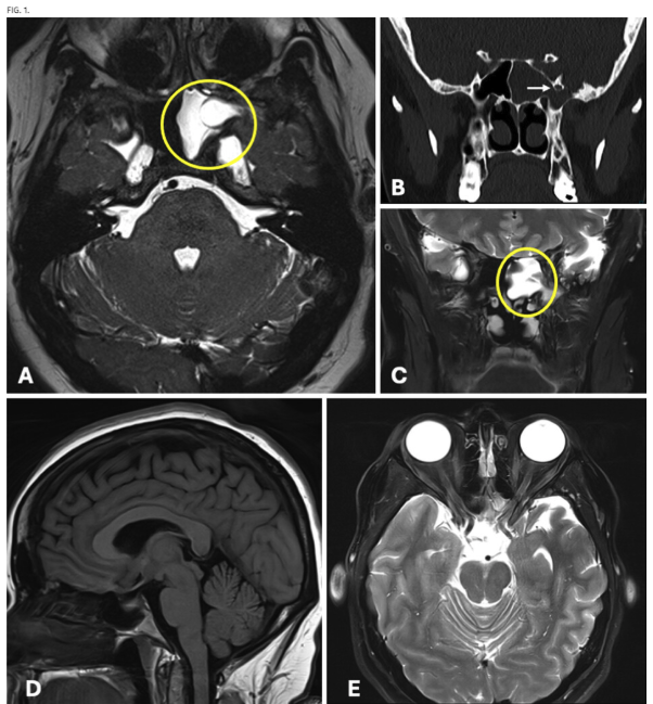
#JNSCaseLessons. Researchers highlight variant case of spontaneous CSF rhinorrhea arising from combined Sternberg canal defect and intracranial hypertension.
https://t.co/702Ph3lSMW
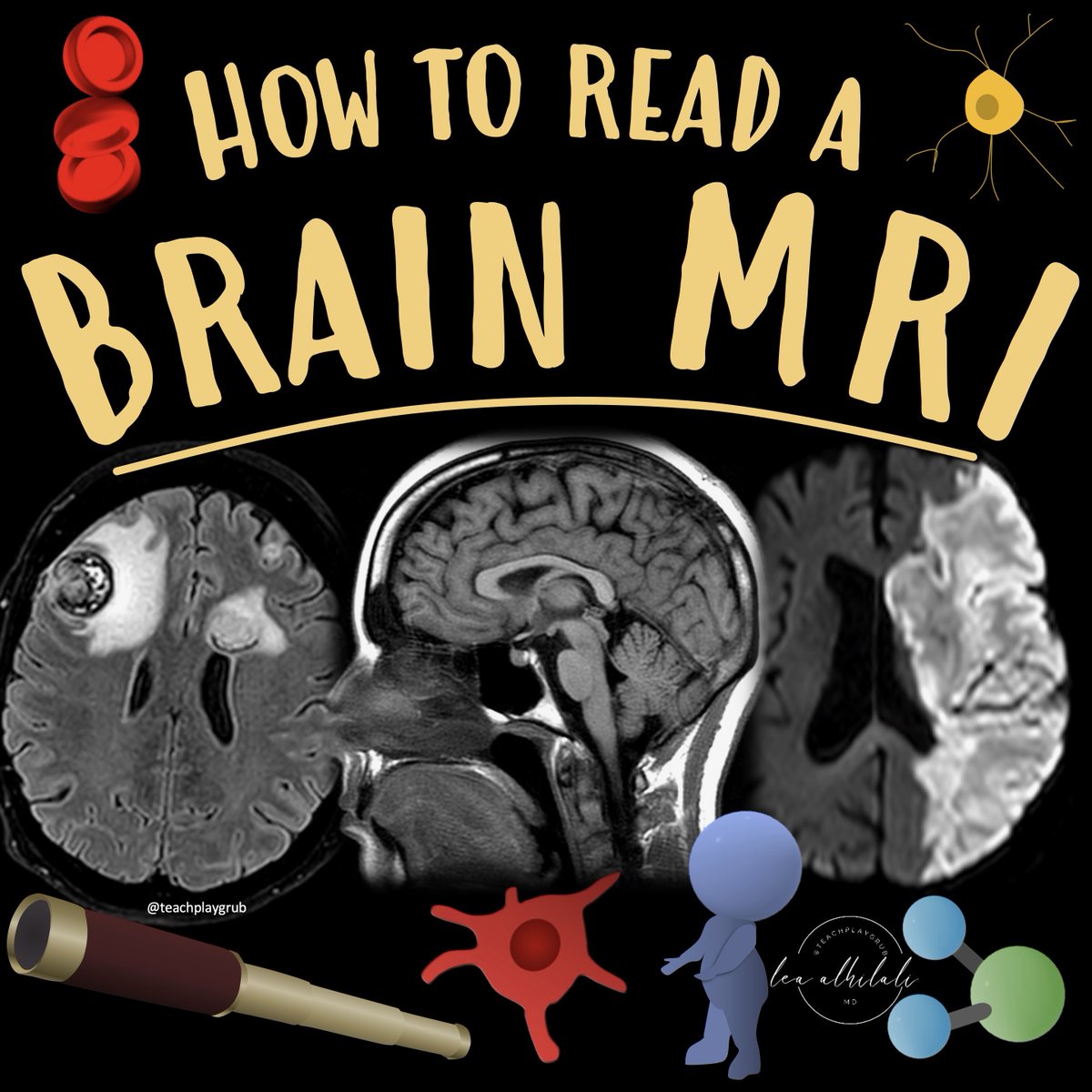
1/Do radiologists sound like they are speaking a different language when they talk about MRI?
T1 shortening what? T2 prolongation who?
Here’s a translation w/an introductory thread to MRI.
#ONSCover Syndromic vs Nonsyndromic Management of Multisuture Craniosynostosis: A Single-Center Experience https://t.co/tW5eoq2CiR by Guillen Arguello et al @LSUHealthNO@cns_update@dgolubMD
#FreeArticle. Researchers discuss clinical scenarios in which shorter fusion constructs may be considered for patients with adult scoliosis.
https://t.co/6SoHlhDthH
Before I had access to nav, I spent much time trying to learn/teach/utilize “freehand” technique. Though I now exclusively employ nav and robotics in my practice, I’m glad I can still rely on these time-tested techniques when needed. Principles first. Adjuncts second.
Spinal cord ependymomas are straightforward to remove when there’s a defined border between tumor capsule and cord, but sometimes they are adherent & difficult to separate from cord, as in this case. The keys: debulk first, collapse capsule inward, work the plane…
The callosal angle in possible iNPH assessment is measured on a coronal plane perpendicular to the anterior commissure-posterior commissure line, at the level of the posterior commissure.
The technique is demonstrated erroneously in this #journalimage
https://t.co/7UAr4QgsHz
1/They say form follows function!
Brain MRI anatomy is best understood in terms of both form & function.
Here’s a short thread to help you to remember important functional brain anatomy--so you truly can clinically correlate!
The spinal cord is a scary thing to incise. The cord has a midline fissure that makes it easier there, but paramedian cord does not. However, myelotomy along dorsal root entry zone (DREZ) is safe for dorsal spinal cord cavernous malformations, entering posteromedial to descending corticospinal tract & dancing around posterior spinal artery…
If you aren’t cheating, you aren’t trying!
Are you looking at MRIs?
Feeling some confusion about the diffusion?
Feel impaired when it comes to the FLAIR?
Wish you could cheat a little?
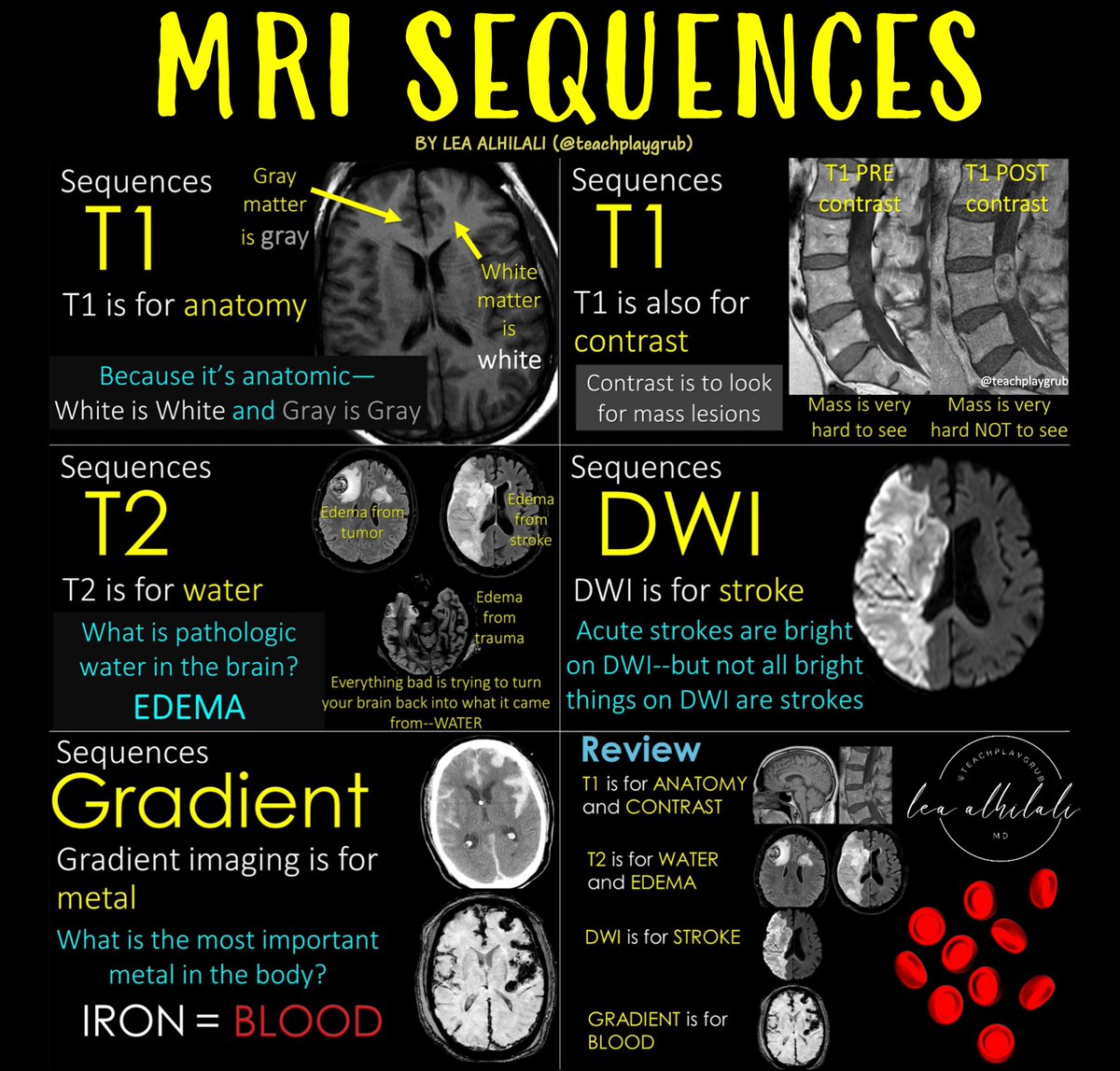
Here’s a post to help you cheat on all the sequences you NEED to know if you are looking at MRIs!
--T1: It’s for anatomy, so brain structures reflect the same color as real life. So gray matter is gray on T1 & white matter is white on T1. It’s also for contrast. Contrast material is taken up by masses making them light up & them easier to see.
--T2: It’s the water sensitive sequence. What is pathologic water in the brain? Edema! My attending once said, “Everything bad in this world is trying to turn you back into what you came from—water."
-- DWI: Diffusion detects stroke, which are bright on diffusion. But anything that makes space tight in the brain can be bright on diffusion (highly cellular masses, clotted blood, pus under pressure, etc.)
--Gradient: Gradient is sensitive to metals. And what’s the most important metal in body? Iron—bc iron is in blood. So gradient is our blood sensitive sequence
So now you know the basic MRI sequences and what they are used for! Now you get rid of all pretenses when it comes to MR sequences!
1/Raise your hand if you’re confused by the BRACHIAL PLEXUS!
I could never seem to remember or understand it—but now I do & I’ll show you how!
A thread so you will never fear brachial plexus anatomy again!