Discover Dermatopathology on the #PathologyPortal! Learn through real cases, digital whole slide images, and expert-led videos by Dr Jerad Gardner, designed for all levels. Topics include tumours, inflammatory conditions, and congenital disorders. Visit:
https://t.co/xtCP7dY4vi
Club de Residentes Iberoamericanos de Cirugía Pediátrica 🌎
Martes 13 de octubre
Invita:
Pontificia Universidad Javeriana, Cali, Colombia
Asociación Iberoamericana de Cirugía Pediátrica
Link: https://t.co/jfjFqYsNT8
Announcing a free @ISGynP sponsored live virtual slide session on Oct 8 @ 12 noon US ET by Dr. Kabbach - "A Practical Approach to Vulovovaginal Non-Squamous Tumors". Register now @ https://t.co/Q2UBdsCCsy
#gynpath#gynaepath#PathTwitter
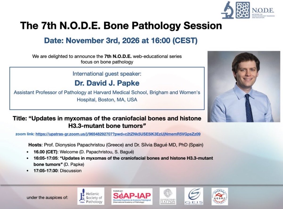
Join us for our next Distinguished Lecture Series session featuring Dr. Jesse K. McKenney
Topic: Spindle Cell Lesions of the Genitourinary Tract
Moderators:Dr. Emily Chan @chemily7, Dr. Rohit Mehra @drmehrarohit
Nov 5, 12 PM Eastern time
Register here: https://t.co/uPaNYi7XxD
Gliomas with pleomorphic and pseudopapillary features (GPAP) are circumscribed tumors with targetable mutations, prolonged survival, and frequent tumor predisposition
https://t.co/7CXOiZspzg
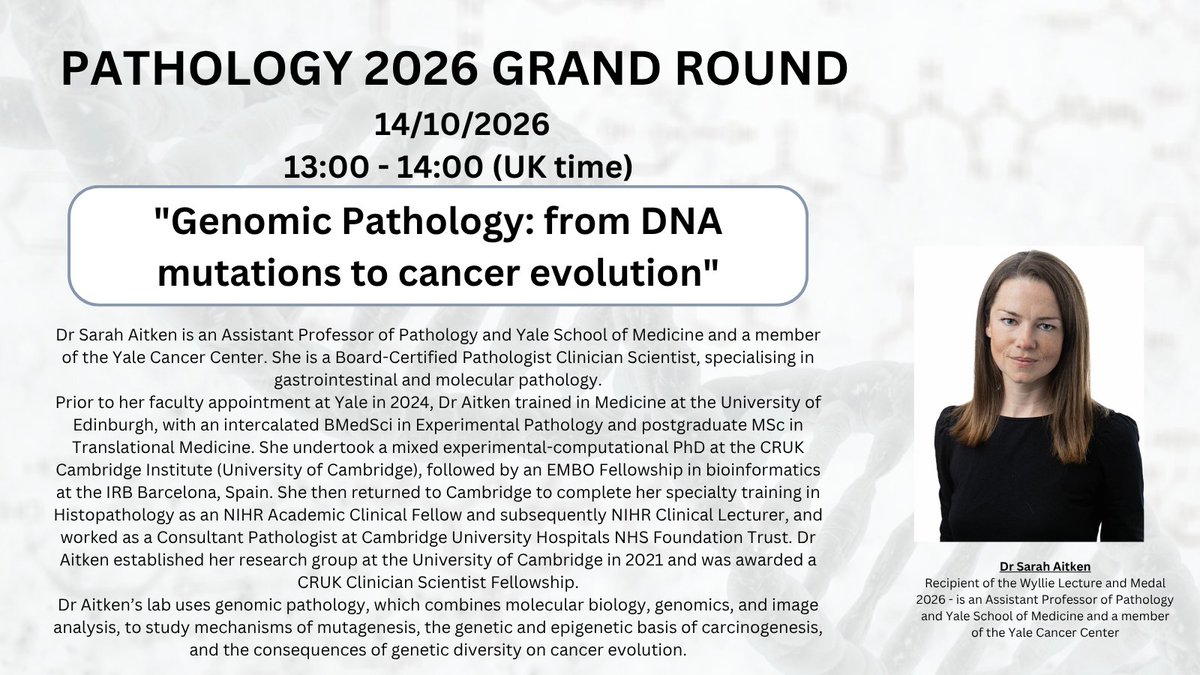
2 WEEKS TO GO UNTIL OUR NEXT PATHOLOGY GRAND ROUND LECTURE!!
Dr Sarah Aitken, recipient of the Wyllie Lecture and Medal 2026, on "Genomic Pathology: from DNA mutations to cancer evolution" 🔬
📅14 October 2026, 13:00 - 14:00 (UK time)
🔗Register: https://t.co/9e8oYyUmrr
Join us on Thursday, Oct. 1 for our next Virtual Education Series: Bridges in Genitourinary Oncology- From Pathology Phenotypes to Clinical Realities.
Speakers: @TingZhaoPathdoc, @DrSwatiBhardwaj, Dr. Gabriela Quiroga-Garza.
🕛 12–1:30 PM EST
Register: https://t.co/tLl1bGJri3
Crowded Gleason pattern 3
Glands are back-to-back and can look cribriform
Pitfall: calling it pattern 4
A keratin stain will highlight the architecture and show they are individual glands
Dr. McKenney. Prostatic Adenocarcinoma Grading: 2026 Update #USCAP#pathology#PathX
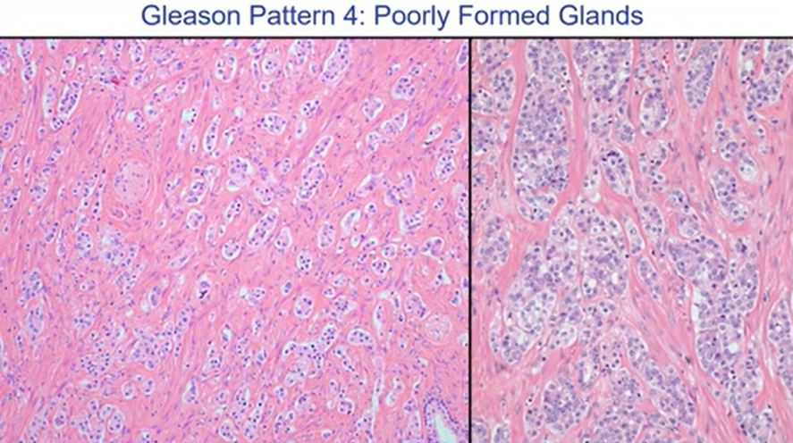
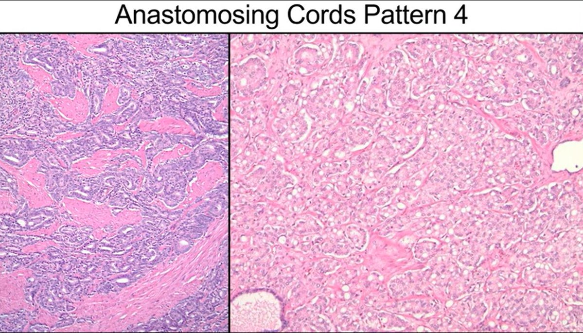
Gleason Pattern 4: Poorly formed glands
None of these glands have lumina, this is not tangential section .
Probably you could think this is grade 5. Do not call it grade 5
Across all clinical outcome groups, this morphologic pattern is NOT associated with metastatic potential.
Dr. McKenney Prostatic Adenocarcinoma Grading: 2026 Update #USCAP #pathX #pathology
Diagnostic Approach to Sinonasal Small Round Blue Cell Tumors
For the initial immunohistochemical evaluation of sinonasal small round blue cell tumors, a screening panel including AE1/AE3, p40, synaptophysin (Syn), SOX10, CD45, CD99, and desmin may be employed to assess epithelial, neuroendocrine, neural crest/melanocytic, hematolymphoid, and myogenic differentiation.
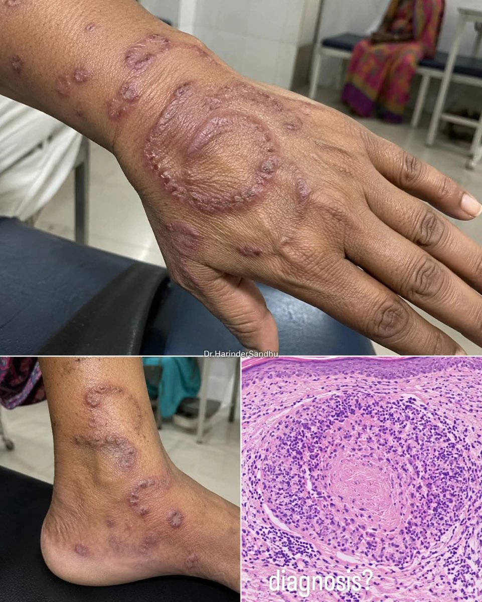
What is your diagnosis for this 45-year-old patient with erythematous-violaceous papules and annular plaques, with biopsy showing neutrophil-rich palisading granulomas surrounding necrobiotic collagen?
Dr. Harinder Sandhu
What is your diagnosis for this patient with multiple erythematous-violaceous nodules and plaques, with histology showing a dense dermal follicle-centered B-cell infiltrate?
Dr. Harinder Sandhu
Prostate - Gleason pattern 3
Well-formed individual glands; some may have a small lumen (pic 1, yellow arrow)
Occasional glands without a lumen and focal branching are acceptable
Evaluate at low power
Dr. McKenney. Prostatic Adenocarcinoma Grading: 2026 Update #USCAP #pathology #pathX
Diagnostic Approach to Large B-Cell Lymphoma
Large B-cell lymphoma (LBCL) is the most common type of aggressive mature B-cell lymphoma. In the 5th edition of the WHO Classification of Haematolymphoid Tumors, LBCL has been further refined into as many as 17 distinct specific subtypes. Cases that do not fit into any defined subtype are classified as diffuse large B-cell lymphoma, not otherwise specified (DLBCL, NOS). The increasing precision of LBCL classification requires pathologists to adopt more accurate and comprehensive diagnostic strategies. Meanwhile, the continuous development of novel therapeutic agents and the diversification of treatment approaches have placed higher demands on precise LBCL subclassification and the identification of clinically relevant molecular alterations.
The diagnostic workup typically includes an immunohistochemical panel consisting of CD20, PAX5, CD3, CD5, CD10, BCL2, BCL6, MUM1, CD138, Cyclin D1, CD30, p53, MYC, Ki-67, and Epstein–Barr virus (EBV)-encoded RNA (EBER) in situ hybridization. In addition, fluorescence in situ hybridization (FISH) analysis is performed to detect genetic rearrangements involving MYC, BCL2, BCL6, and IRF4, among other relevant molecular abnormalities.