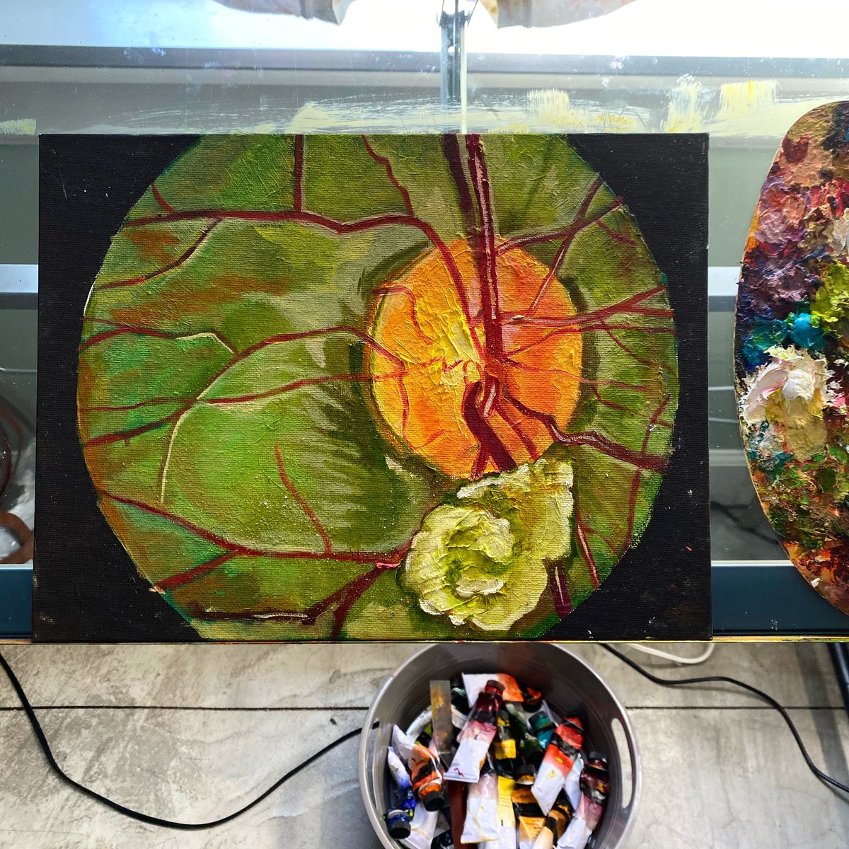
I made a larger version of my ARMD w/ geographic atrophy piece for a client a little while back. It was inspired by a photograph with such flamboyant colors. If you come across fundus photos with colors as vibrant as this, feel free to send them my way.
Fundus portraits I made for a client. This person had pseudo-papilledema and fish drusen (swipe right to see the photographs I was sent) Did I do them justice???
Eye docs compliment my work but one thing I wish I got from them is more constructive criticism. Am I doing the ocular pathology justice? Are there certain things I need pay closer attention to? Is there aspects of fundus anatomy that I could portray better in my art?
Excited to share this unique appearance of macular myelinated retinal nerve fibre layer published in @AAOjournal
MRNFL is normally associated with a benign outlook but here we show that anatomical location of the MRNFL can have an impact on vision.
https://t.co/oH3CMAFzAp
I made a larger version of my ARMD w/ geographic atrophy piece for a client a little while back. It was inspired by a photograph with such flamboyant colors. If you come across fundus photos with colors as vibrant as this, feel free to send them my way.
A picture of an #eye after #surgery of a #retinaldetachment operated within less than 24 hours of presentation to fix the detachment and restore #vision. This was a picture taken with around 50% gas #tamponade fill!
I have been chuffed to see a glad pati…https://t.co/jAIkis2ziP