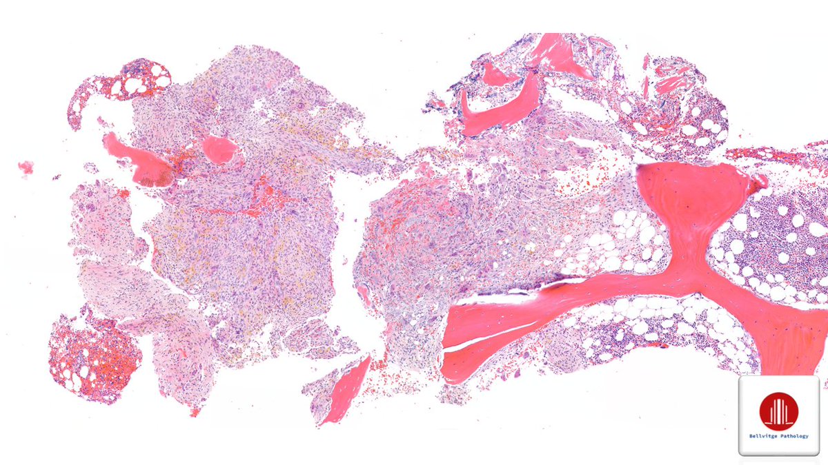
One image of this cerebellar tumor is all you need. But if you were to do just one immunostain, what would it be? (Don't be INHIBIted, give it your best guess.) #pathology#neuropath#pathtwitter
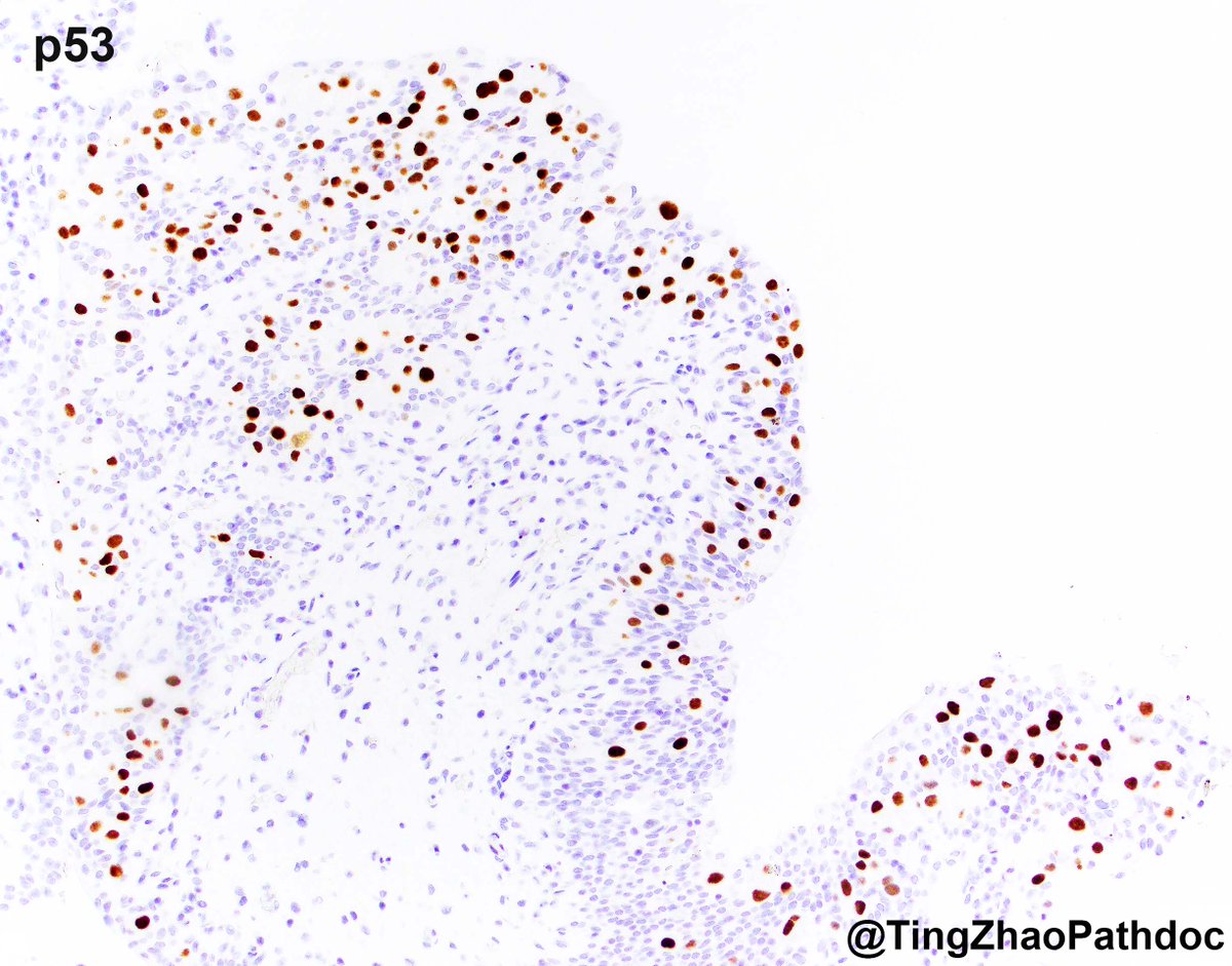
Urothelial carcinoma in situ (UCIS) with plasmacytoid features #GUpath
➡️UCIS with cellular rounding, enlarged eccentric nuclei, and dense eosinophilic cytoplasm
➡️Diffuse/strong CK20 in ~95%, mutant p53 (overexpression or null) in ~35%, absence of CD44 reactivity in the neoplastic cells in ~65% (PMID: 31368912)
➡️In Dr. Sangoi’s case series, none of the 23 plasmacytoid UCIS are associated with the plasmacytoid subtype of invasive urothelial carcinoma (PMID: 31368912) @slusagar
➡️One study reported a plasmacytoid invasive urothelial carcinoma associated with plasmacytoid UCIS (PMID: 18379419)
➡️This patient’s TURBT prior to cystectomy showed invasive urothelial carcinoma with extensive (>90%) plasmacytoid features
➡️Different morphological patterns of UCIS does not appear to impact prognosis (PMID: 23397277)
CD70/CD27 signaling may be a key driver of myeloma progression — especially extramedullary disease — and a druggable target.
https://t.co/3u5zw38KiN
What they found:
• CD70-high myeloma = worse OS
• • CD70/CD27 activates MAPK/ERK + Wnt → drives proliferation
• ADCC-enhanced anti-CD70 (cusatuzumab) shut down tumor growth in xenograft models
#Myeloma Paper of the Day: CD70/CD27 signaling promotes pathogenesis of myeloma & represents a promising therapeutic target w/ ADCC-enhanced anti-CD70 antibody, cusatuzumab, which demonstrated high efficacy vs. myeloma in xenotransplantation models: https://t.co/qToX27gs9i. #mmsm
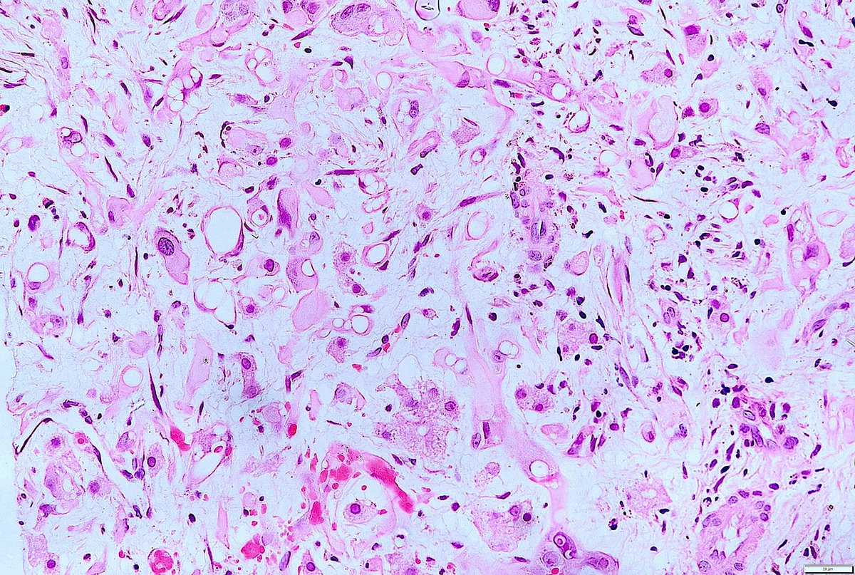
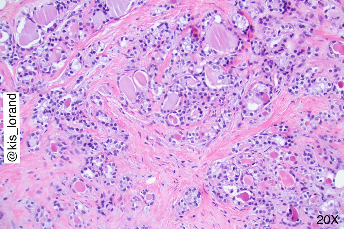
A rare primary #tumor of the #liver: Calcifying nested stromal epithelial tumor. Well defined nests of spindled to epithelioid cells surrounded by desmoplastic stroma. #Psammoma-like #calcifications & #osteoid material may be present. AE1/AE3, WT1 & abnormal b-catenin (+).
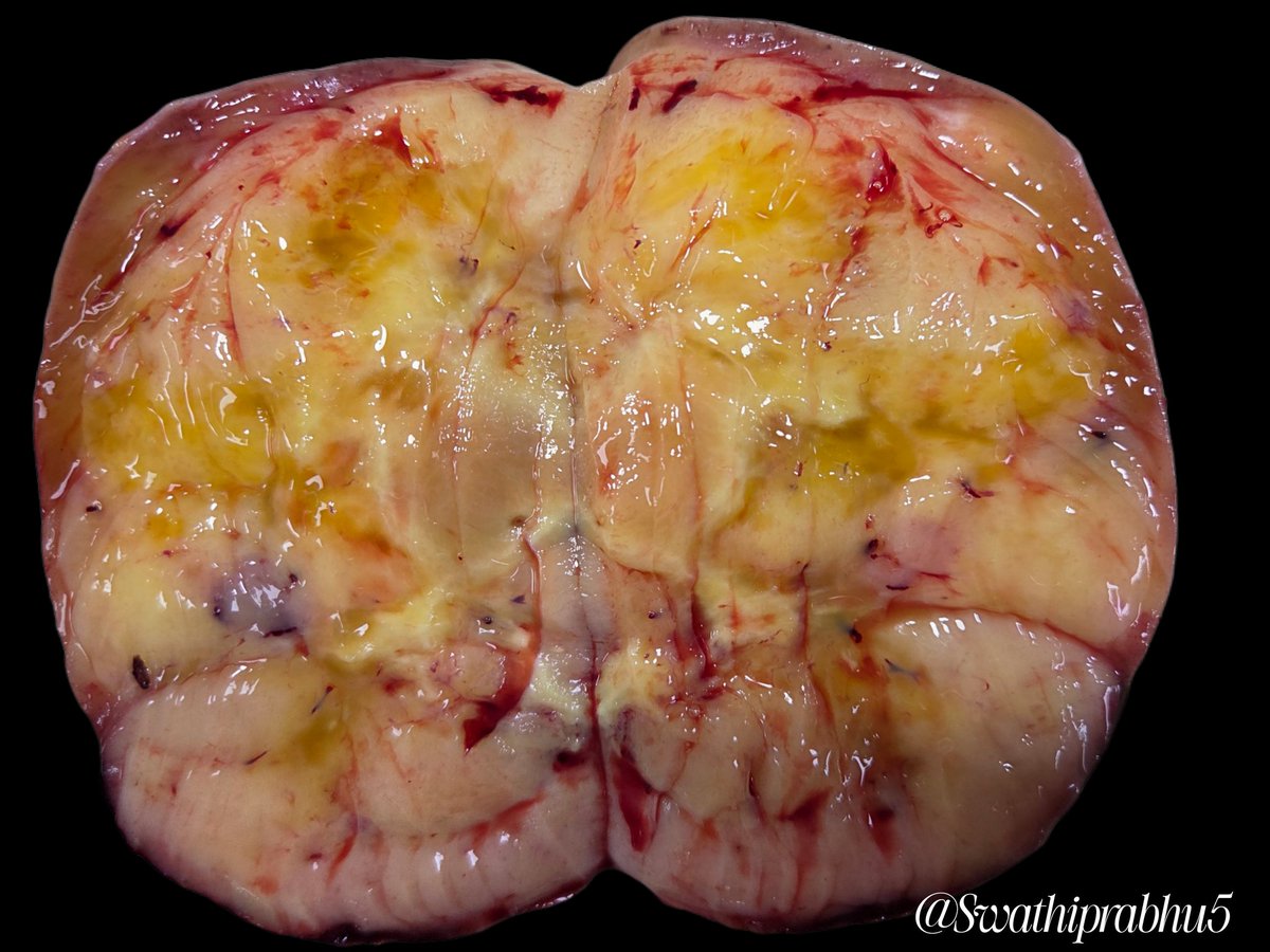
So it all fits: the bone lesion is a nice example of 𝐛𝐫𝐨𝐰𝐧 𝐭𝐮𝐦𝐨𝐫 (𝐠𝐢𝐚𝐧𝐭 𝐜𝐞𝐥𝐥 𝐫𝐢𝐜𝐡 𝐥𝐞𝐬𝐢𝐨𝐧) 𝐢𝐧 𝐭𝐡𝐞 𝐬𝐞𝐭𝐭𝐢𝐧𝐠 𝐨𝐟 𝐩𝐫𝐢𝐦𝐚𝐫𝐲 𝐡𝐲𝐩𝐞𝐫𝐩𝐚𝐫𝐚𝐭𝐡𝐲𝐫𝐨𝐢𝐝𝐢𝐬𝐦, "brown" due to the presence of hemosiderin.
Breast biopsy with the following histology. Classical 3 questions 😀
1. What is the phenotype of the cells?
2. What is your understanding about the GATA3 expression of the cells?
3. If you see the same lesion in cervical biopsy what would you call it?
#pathology#breastpath
#Myeloma Paper of the Day: Spatial transcriptomics reveals profound subclonal heterogeneity and T-cell dysfunction (exhausted TIM3+/PD-1+ T-cells) in extramedullary myeloma with oxidative phosphorylation representing the major metabolic program: https://t.co/DhA5WYmVKY. #mmsm
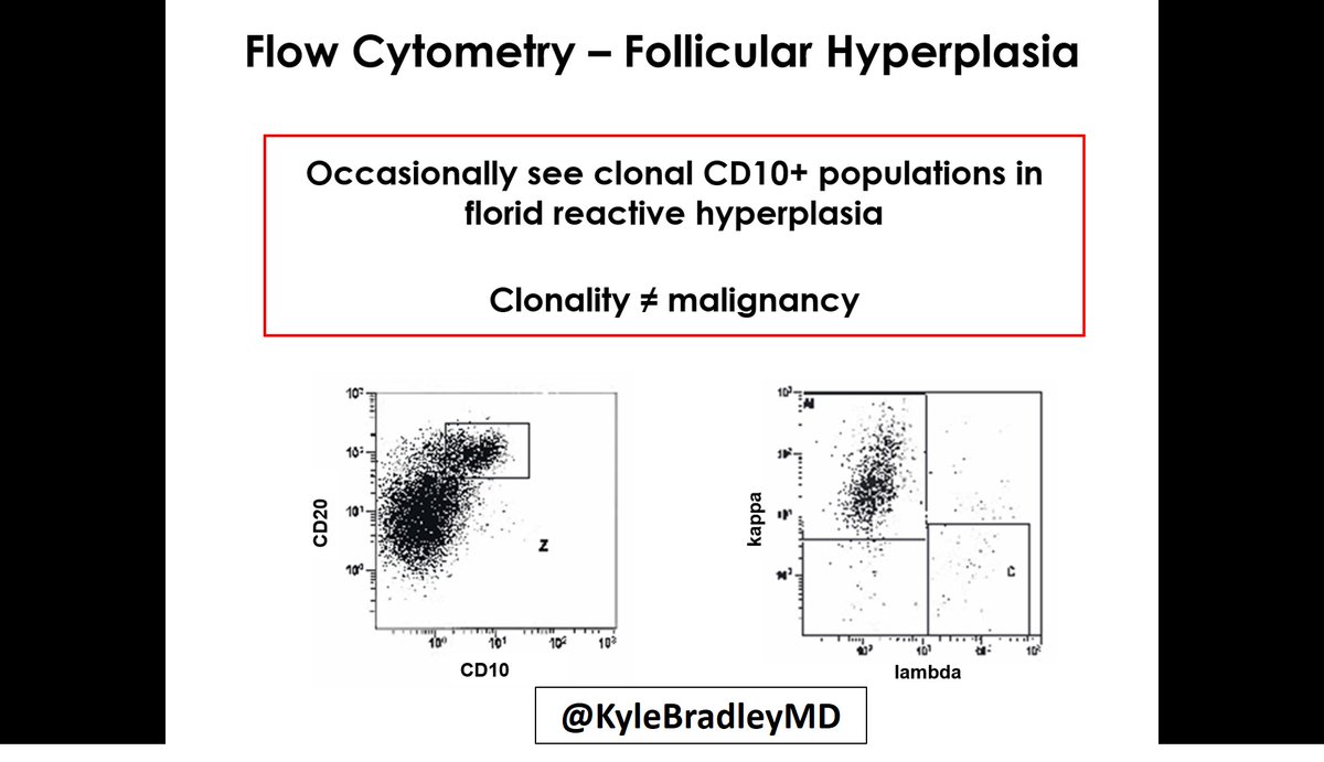
Flow cytometry pitfall:
Some cases of reactive florid follicular hyperplasia will have a clonal B-cell population.
Usually CD10+/CD20 bright.
Background polytypic B cells are usually present.
#CAPVirtualPath