@eddyjoemd Not routinely, but in selected patients—particularly when high airway pressures and hypoxemia are a concern in the setting of altered chest wall or abdominal compliance.
Also, quite expensive over here.
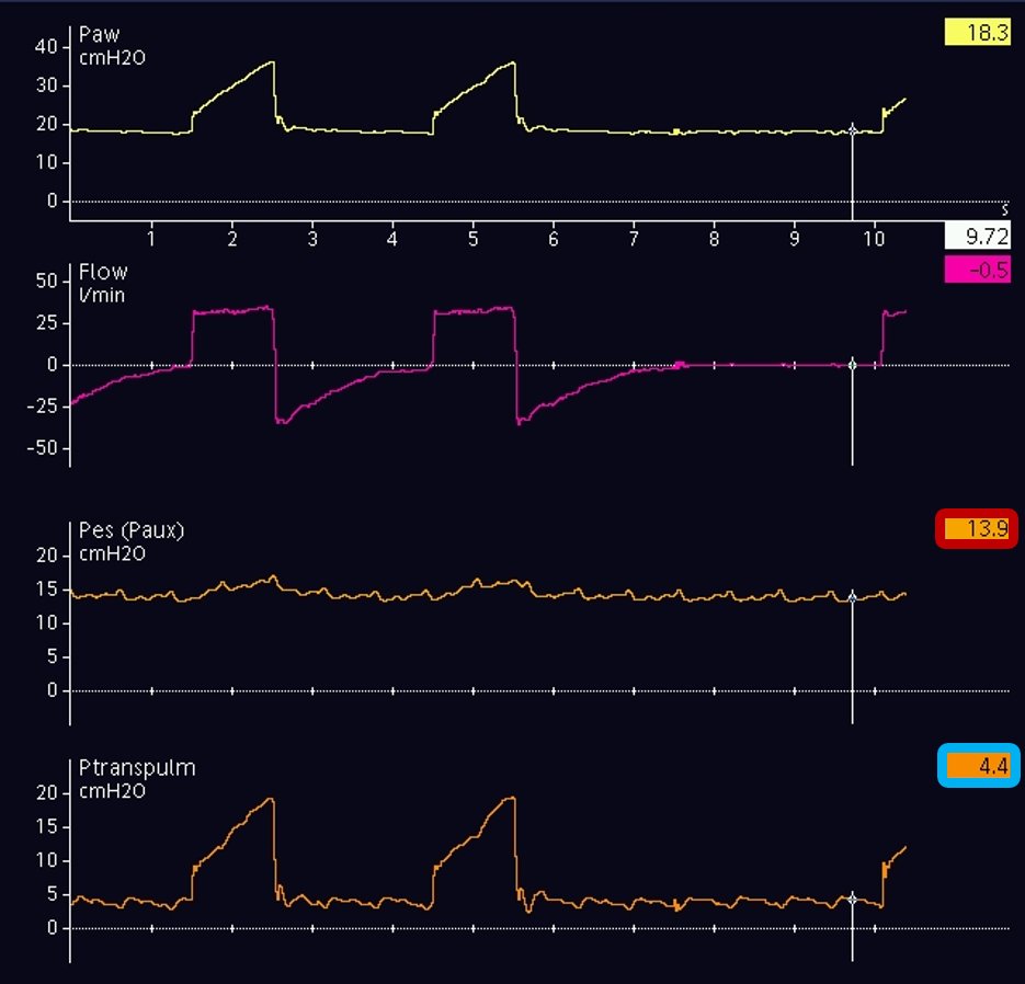
Post-arrest aspiration pneumonitis in an obese patient. EPM placed and PEEP titrated to 18 for an end exp TP of 0 (Fig 1).
In 15 minutes, end exp TP is +4 w/o setting changes (Fig 2).
What happened? Poll and answer below! Repost if you learn something new!
@ross_prager Hey Dr Prager! Recently reading through some cases on this topic... What's your opinion about early plasmapheresis with severe shocky patients, particularly with cardiogenic shock?
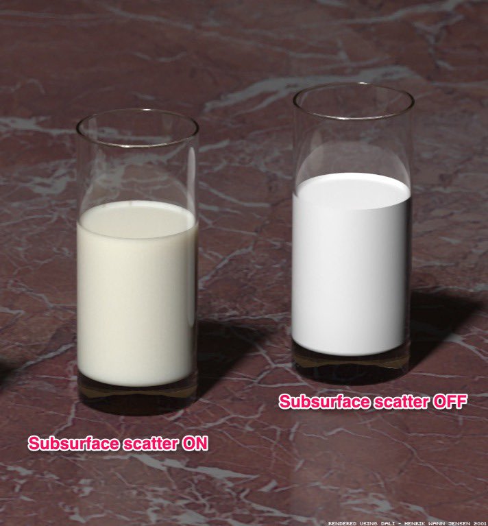
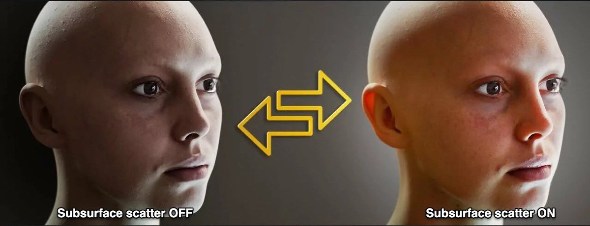
Sub surface scatter is present in lots of materials (milk, wax, marble, etc) but it’s especially important for living things.
It turns out that the movement of blood through capillaries* is a major contributor to SSS in living things.
(Normally blood flows ~1 mm/sec in skin)
5/
Dr. Pablo Cruces, pediatric intensivist at El Carmen Hospital (Chile) and translational medicine guru, shows us how P-SILI concentrates in the juxtadiaphragmatic regions of the lung, with force vectors directed outward at the pulmonary capillaries.
Heart, Lung & Critical Care 26'
Loss of pulsatility after #ECPR?
🔎 221 pts (2011 - 2024), high volume ECMO center
🖥️ loss of pulsatility = any documented PP = 0 during first 12 h after #ECMO
🖥️ at least 1️⃣ episode occurred in nearly 50% of patients. They had:
⚖️ higher proportion of #OHCA + longer CA-to-ECMO
⚖️ higher in-hospital mortality; each additional hour of loss of pulsatility increased risk of death
#FOAMcc #FOAMecmo @Resus_Plus
🔓 https://t.co/btjmx1XL1x
#POCUS#Nephpearls#MedEd 📑
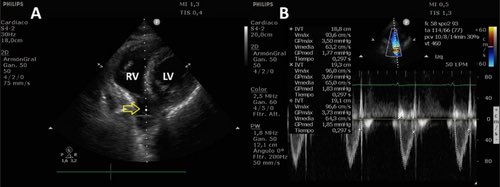
Correlation Between RVOT VTI Measured via a Modified Subcostal View and LVOT VTI in Critically Ill Patients.
“An RVOT VTI > 14.6 cm predicted LVOT VTI ≥ 17 cm with 80.7% sensitivity (95% CI 62.5%–92.5%) and 100% specificity (95% CI 73.5%–100%).”
- by @Issac80 and team
🔗 https://t.co/a6kinNW9sN
Critical Physiology Series #25
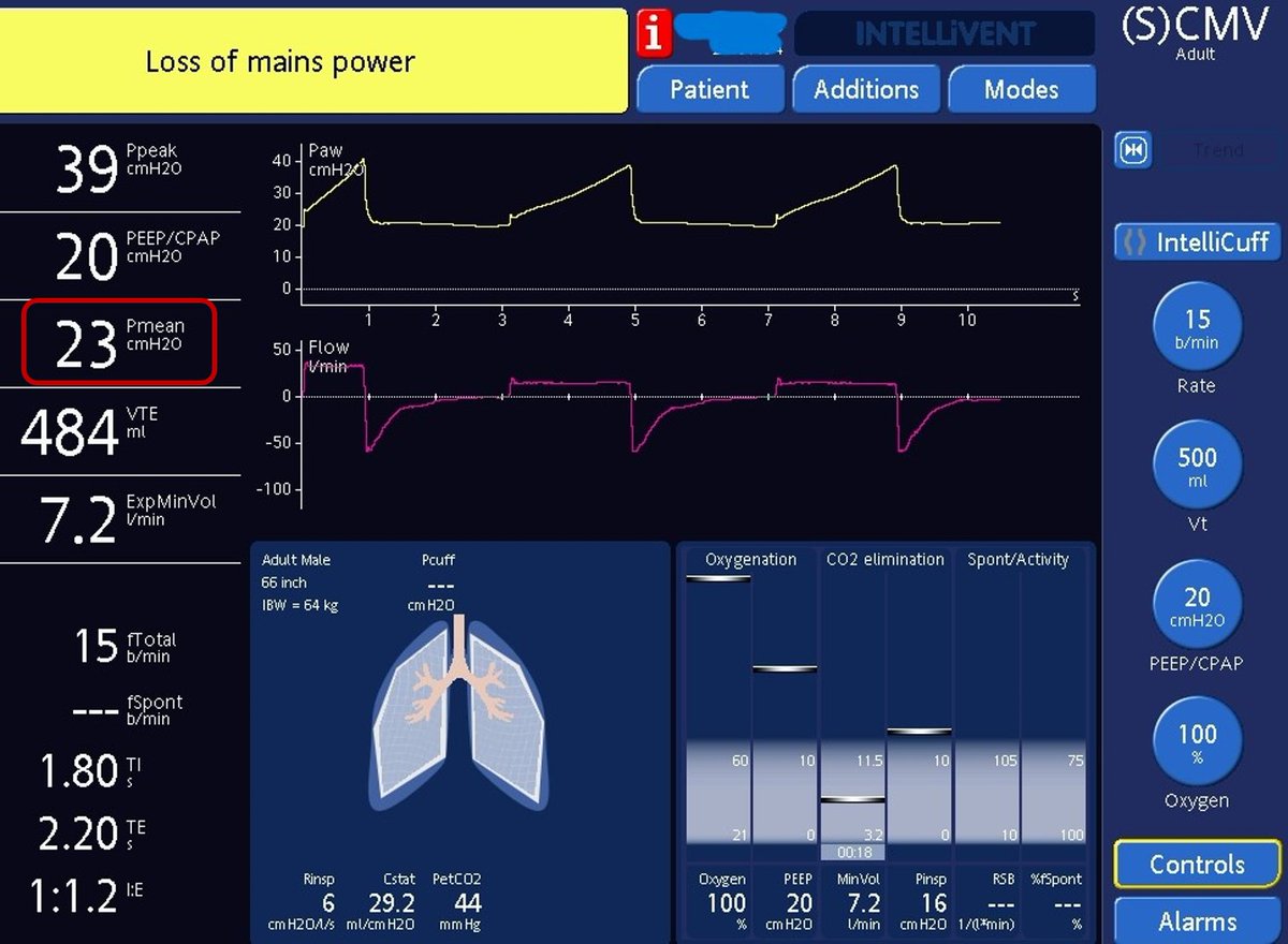
Pmean, Compliance and Transpulmonary Pressure During Mechanical Ventilation
Mean airway pressure, or Pmean, is the average pressure applied at the airway opening during the complete respiratory cycle.
It is influenced by PEEP, inspiratory pressure, inspiratory time, respiratory rate and the pressure waveform. Increasing Pmean may improve alveolar recruitment and oxygenation, but it may also increase intrathoracic pressure, reduce venous return and increase right ventricular load.
Compliance describes how easily the respiratory system expands.
Static compliance is calculated using tidal volume divided by plateau pressure minus total PEEP. Because it is measured during an inspiratory pause when airflow has stopped, it mainly reflects the elastic behaviour of the lungs and chest wall.
Dynamic compliance uses peak inspiratory pressure instead of plateau pressure. It therefore includes both elastic load and airway resistance.
When dynamic compliance falls but static compliance remains stable, the problem may be increased resistance from bronchospasm, secretions or an obstructed tube.
When both fall, reduced lung or chest wall compliance is more likely.
However, airway pressure does not act only on the lungs.
Part of it expands the chest wall. Transpulmonary pressure is the difference between airway pressure and pleural pressure and therefore estimates the pressure actually distending the lung.
Pleural pressure can be approximated using an oesophageal balloon.
This distinction is important in obesity, abdominal hypertension, chest wall stiffness and severe ARDS. A high plateau pressure may partly reflect a stiff chest wall rather than excessive lung stress. Conversely, strong spontaneous inspiratory effort may create a dangerously high transpulmonary pressure despite an apparently acceptable airway pressure.
The bedside message is simple.
Pmean describes average airway pressure over time.
Static compliance describes elastic behaviour.
Dynamic compliance includes resistance.
Transpulmonary pressure separates lung stress from chest wall pressure.
Safe ventilation requires understanding where the pressure is going, not only what the ventilator displays.
Recommended lectures 📚
Hess, 2014, https://t.co/LtGTXmTeF3
Placenti, 2024, https://t.co/X6Phljyqfh
Grieco, 2017, https://t.co/fPPIVy2mwz
@franciscojlk Great post!
It reminded me of this outstanding review. It explores several additional physiological variables that can help us truly implement a multimodal lung- and diaphragm-protective ventilation strategy during weaning. Highly recommended.
https://t.co/TOnH87e94p
As an ER-trained intensivist, I still find it difficult to completely let go of therapeutic hypothermia after cardiac arrest. The evidence has evolved, but so has our understanding of its limitations. Whatever our current practice, we stand on the shoulders of giants!
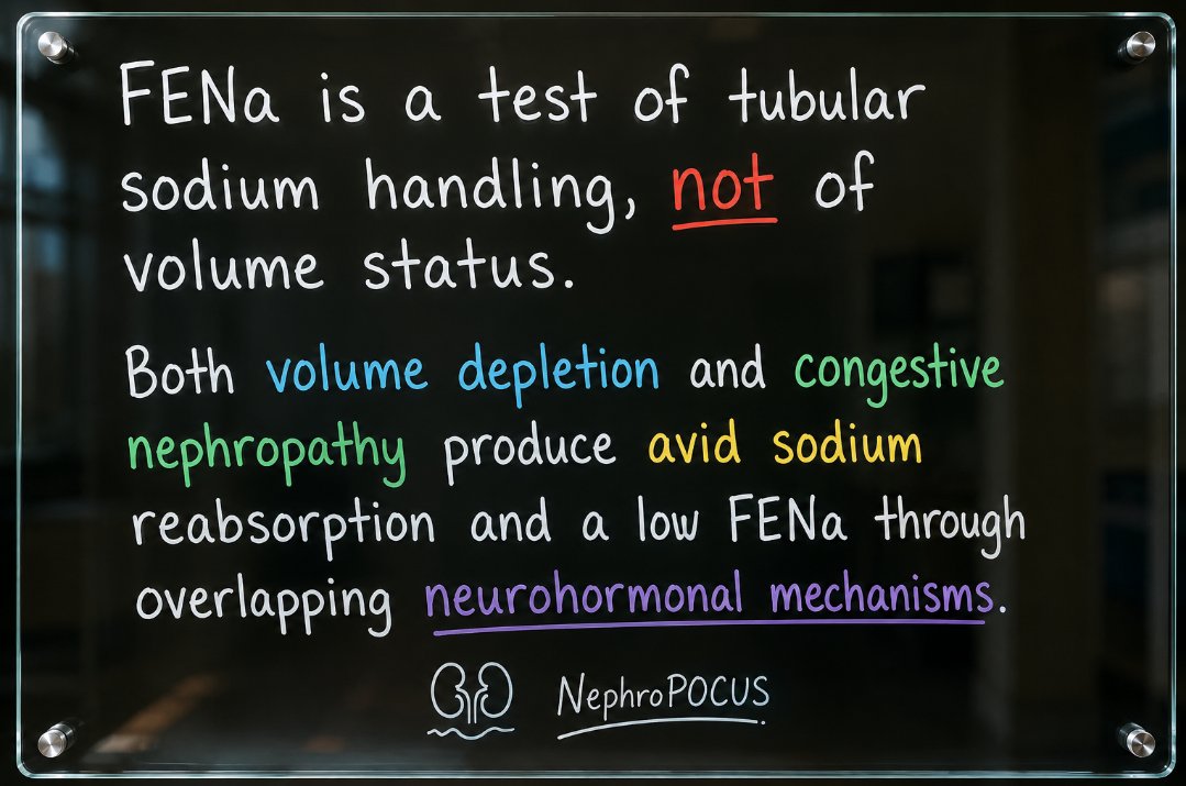
Somewhere, some doc 👨⚕️: "Urine sodium is low in this patient with #AKI. Must be pre-renal AKI. Let's give fluids."
Meanwhile the physical exam (#POCUS)👇
#Nephpearls#FOAMed
Answer: Add end-inspiratory pause!
The end-inspiratory pause is a setting that efficiently increases mean airway pressures. Depending on the ventilator, itime is either directly set by the user so preset inspiratory flows are a result of Vt and itime, or are indirectly set by the user choosing insp flows and Vt. For example, if I choose 1L Vt and flows of 1L/min (i.e., 60L/min), itime would be 1s. To get to an itime of 1.5 seconds, assuming constant flows, I would need to lower flows to 45L/min.
Although going up on the itime knob or lowering preset inspiratory flows will accomplish the goal of extending the duration of inspiration, low flows will lead to slow pressurization of the lungs. Therefore, although you are prolonging itime, you are not efficiently increasing mean Paw.
By doing an end-inspiratory pause with each breath, you get quickly to your end inspiratory pressures and extend that pressure for the desired duration of inspiration.
Note in the waveforms below how mean airway pressures are higher (in red) when itime was extended with an insp pause (left) versus with prolongation of itime/lowering of inspiratory flows (right).
#echofirst
✅ Time to close w/🙏 all comments
✅ Answer: Localized RV & LV tamponade due to LV perforation & pericardial hematoma (following a ViV MVR, TTE apical 4 chamber view shows bioprosthetic MVR & large atria)
✅ Apical echo free space was new compared to pre-procedural TTE (image 1 ⬇️)
✅ Diagnosis of perf was quickly confirmed by use of UEA (Image 2 ⬇️)- following LV opacification contrast was seen in pericardial space within a few seconds (Apical 4Ch view)
✅ Chest CT also showed the pericardial fluid (image 3 ⬇️)
✅ LV perf is rare (<1%) after transcatheter MVR or TEER
✅ Preferred Rx is surgical when tamponade is present
✅ No aortic gradient present despite the 2D proximity of MVR to septum
@DrRajeshG1@argulian@EchoProsto@BryanTanMD@AnkurKalraMD@sudarshanballa@iamritu@Hragy
Clinical Question :
When managing Paroxysmal Supraventricular Tachycardia (PSVT), we routinely utilize a 3-way stopcock to perform the two-syringe technique for Adenosine administration, as demonstrated in the video..
Why must Adenosine be administered using the rapid two-syringe technique (drug + 20 mL saline flush)? And what physical maneuver should be performed on the patient's arm immediately after injection to enhance efficacy?🤔🤔
@DanRozenbaum Thanks for the post Dr Rosenbaum! Awesome as usual.
One question: why was the I:E ratio in Pic1 2.5:1 in an obstructive physiollogy patient?