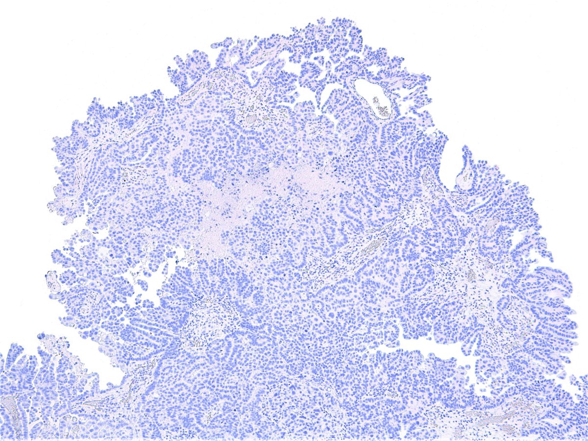
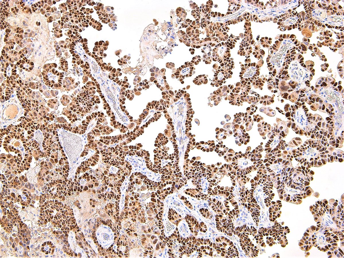
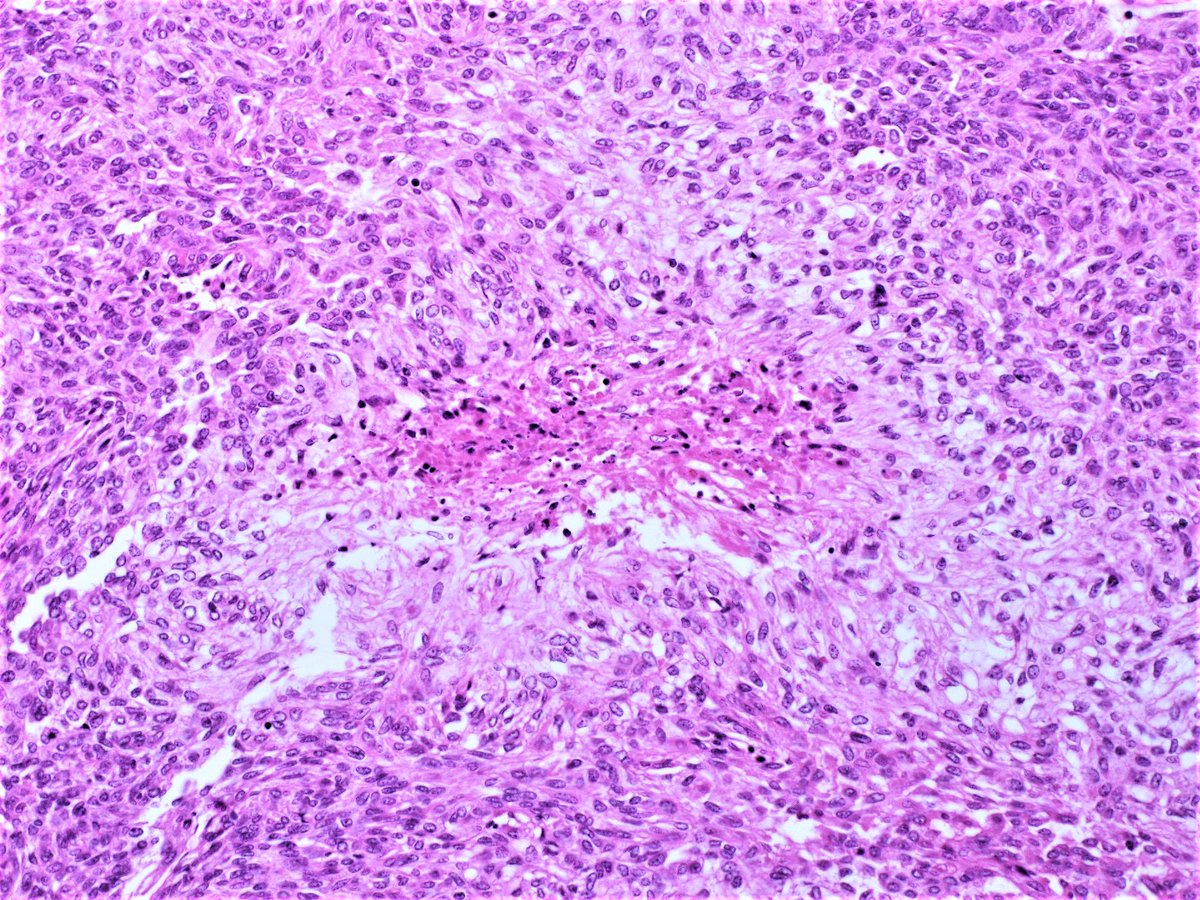
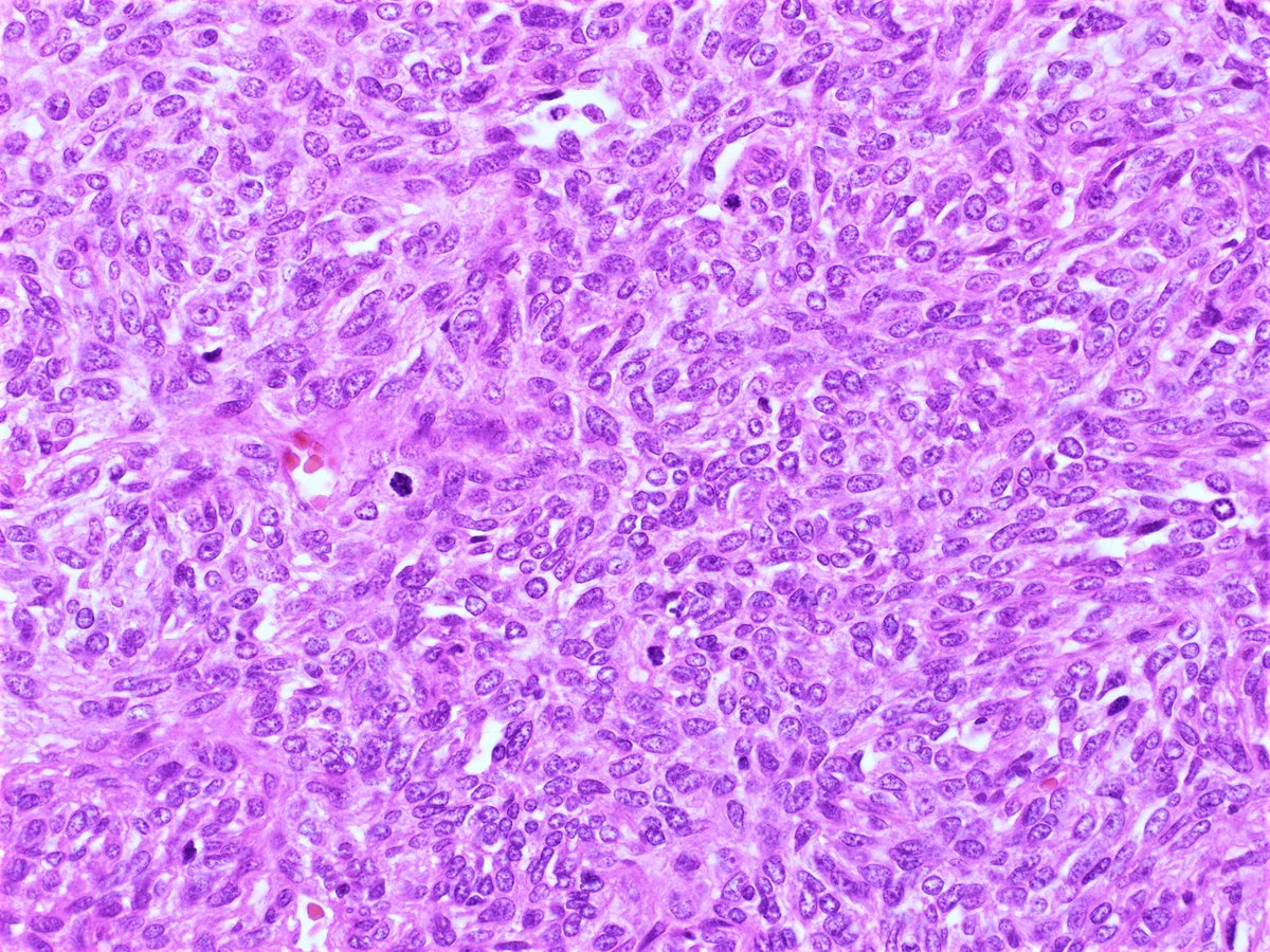
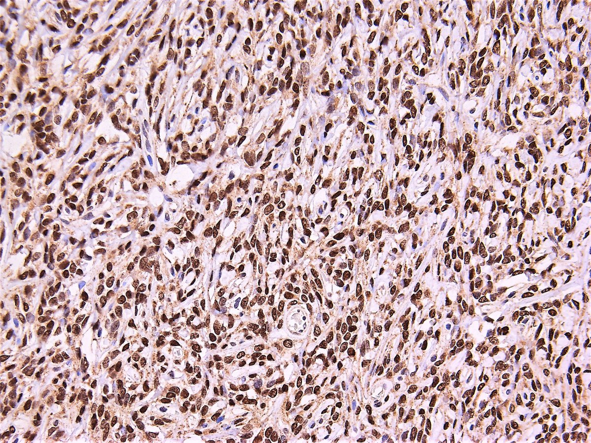
Immunostains, HNF-1beta, p53, Vimentin, p16INK4A, respectively.
My final diagnosis is clear cell carcinoma of uterine corpus. However, I understand that there may be different opinions.#pathology#Gynpath
https://t.co/yjjJndzgUm
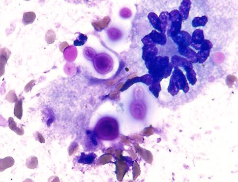
Seminoma:
-Large dispersed cells
-Voluminous cytoplasm with peripherally located vacuoles of glycogen (blister like cells)
-Macronucleoli
-Background of lymphocytes
-Granulomatous inflammation
@roula_katerji@catherinejroe @dannyjlubinpath @vaipath@ac_pathgal@DrGeeONE
I cannot overstate more, the importance of having a significantly higher threshold for a Papillary Thyroid Carcinoma (PTC) diagnosis in the presence of Hashimoto thyroiditis (HT) on FNA. This case however, did turn out to be multifocal PTC arising in an exuberant HT.
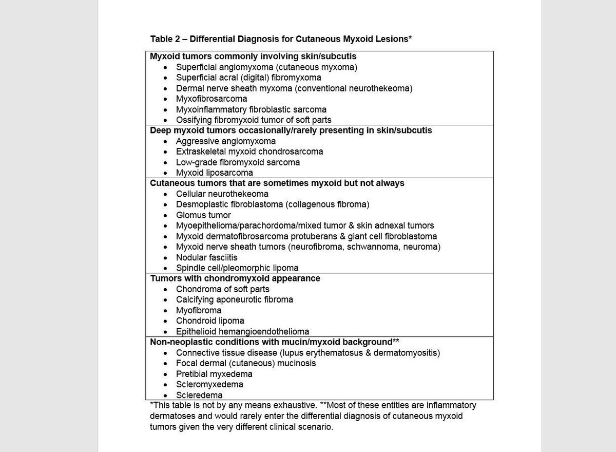
Confused by myxoid tumors? Then check out this myxoid review article by me & @NRiddleMD. Download free here: https://t.co/xK9l7h7Ya1. See also my myxoid video playlist: https://t.co/HH6iusjPRx #pathology#dermpath#BSTpath
When "AGE" of the patient may influence your judgement. In Thyroid FNA of a 14/M, you expect to see Papillary or Medullary CA but NOT a Hurthle cell neoplasm. The resection on this showed Hurthle Cell Carcinoma (quite rare at this age).
A beautiful example of “hypervacuolization” of malignant epithelial cells in a “Papillary Thyroid Carcinoma, Cystic Variant”. Don’t disregard them as simply histiocytic.
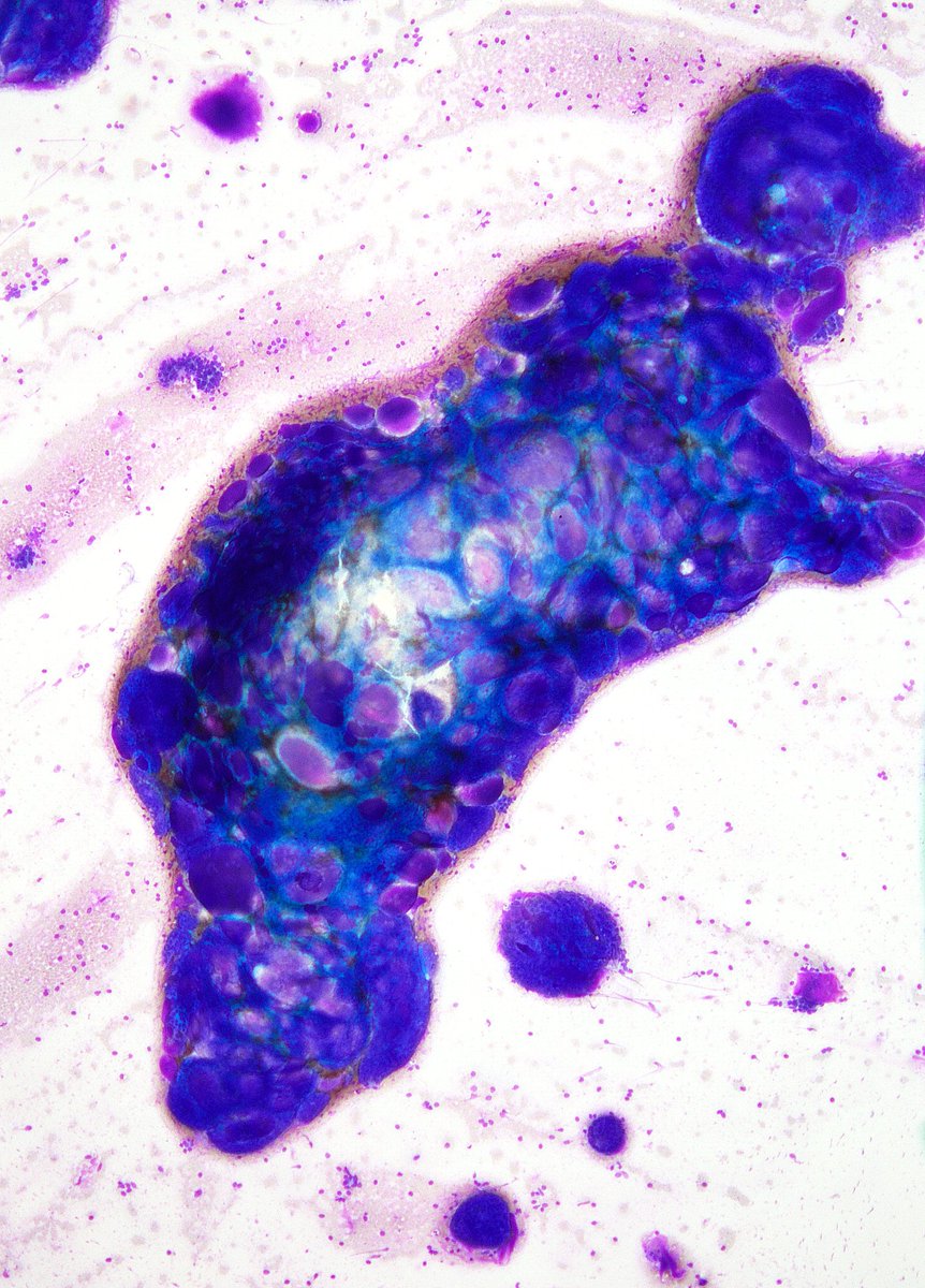
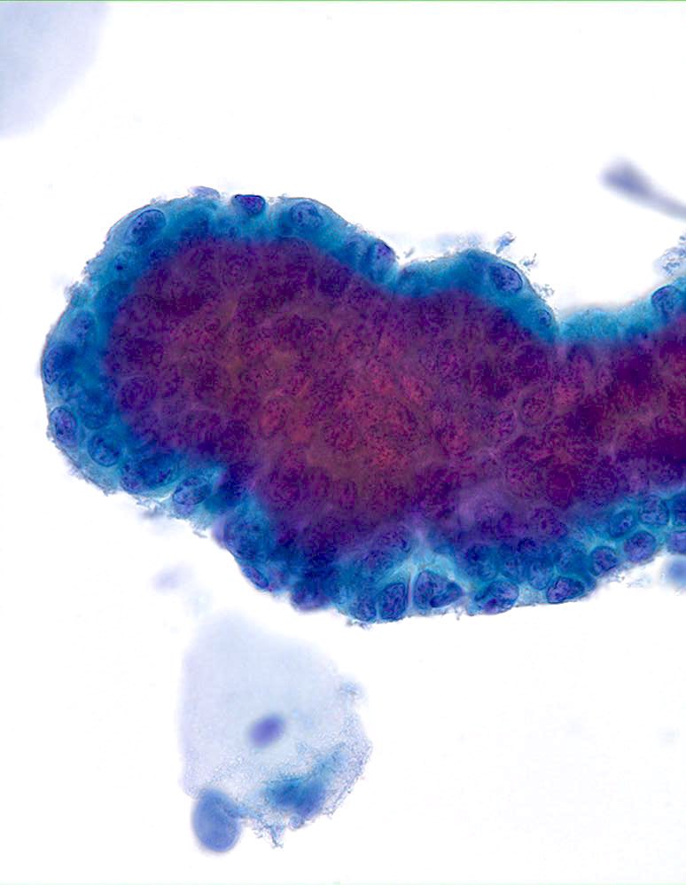
The FORMIDABLE world of PAP Test Cytology. How high can one go on this scary-looking “Glandular” fragment? It was called “AGC-EC”. Follow-up showed an “Endocervical Polyp”. (ThinPrep)