Registration is now open for the 2022 GAP Annual Meeting!
✳️ Join us Oct 28-29th in Atlanta, GA ✳️
Learn more & register at the link below:
https://t.co/oOLX9vaBZp
We look forward to seeing you this fall! 🎃
#GAPath2022@EmoryPathology @AU_Pathology
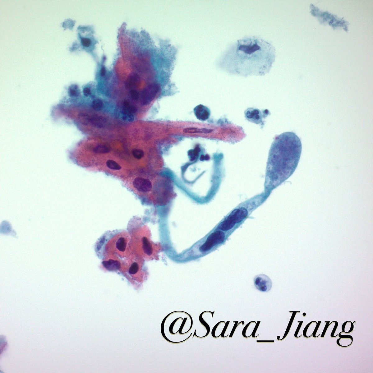
Glomus tumor – FNA of one of many reportedly painful foot nodules shows uniform round cells lining branching capillary-sized vessels. Cytology on these sometimes just shows round cell clusters, but this case had nice architecture on smears. SMA+ cute pericytic neoplasm.
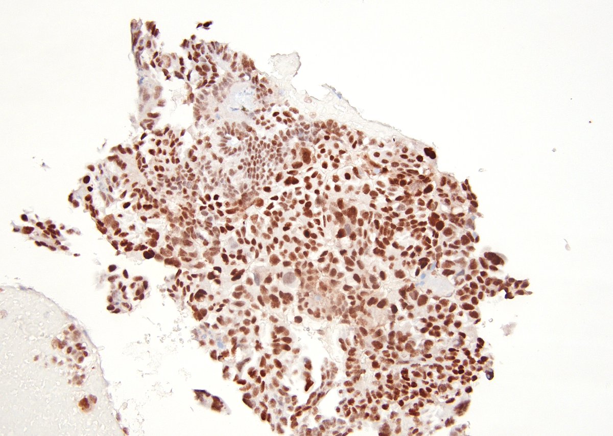
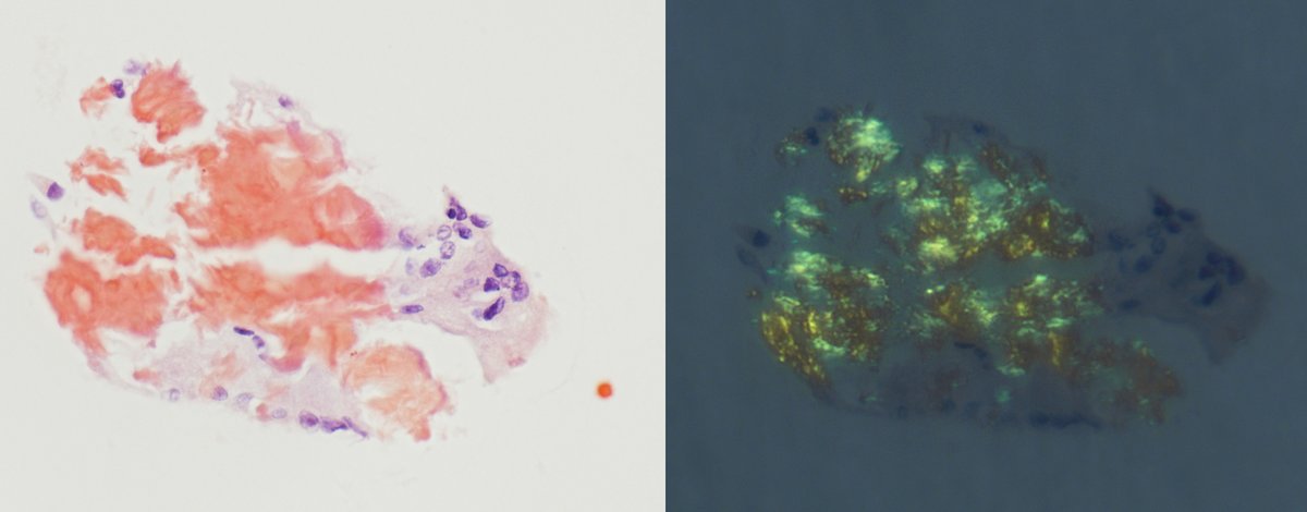
Medullary thyroid carcinoma – hypercellular FNA of mostly dyshesive, plasmacytoid cells w/ neuroendocrine chromatin, red granules (arrow), and admixed amorphous material. Calcitonin+/chromo+ and congo red+ amyloid w/ apple green birefringence (shown). Can trigger workup for MEN2.
We have an unexpected Cytopathology Fellowship position available for this coming academic year 2022-2023!
Come join our fam in ATL!
Email [email protected] for information.
https://t.co/cCXSMbPAzW
#pathtwitter#cytopath#cytology
A visually striking composition of branching vessels and background mucin from a breast FNA. A small group of carcinoma cells @ the bottom right. #cytopath#cytology#FNA
REPOST of Emory Case of the Week #17
.
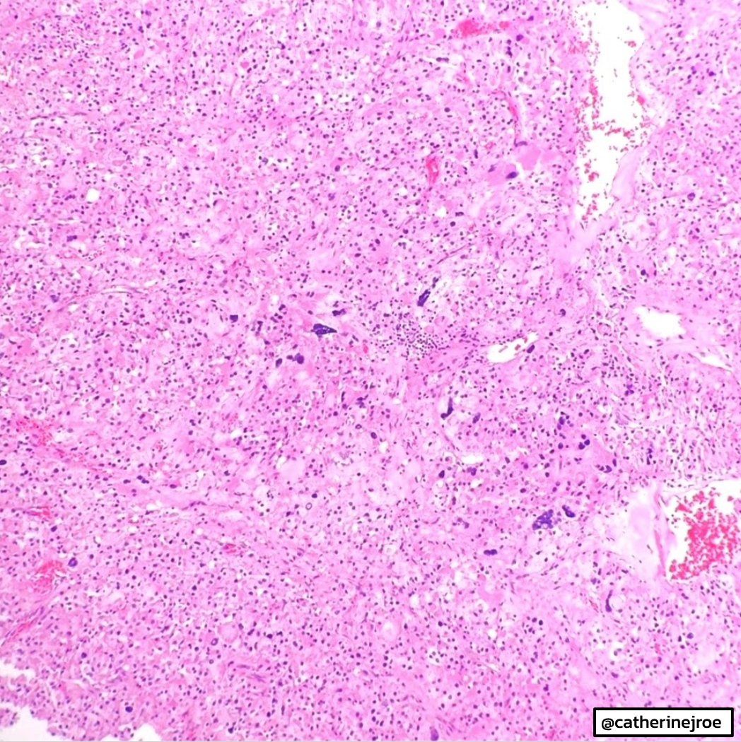
54 y/o female presents with 12 cm adrenal mass. Radical nephrectomy performed.
Answer the question below!
Contributor: @catherinejroe#EmoryCoTW#gupath#endopath
Complete mole
>>Diffuse, villous enlargement with marked hydropic changes
Cistern formation and trophoblastic hyperplasia in a circumferential pattern
>Cytologic atypia
>Broken strands of fibrillar material (cistern formation)
p57 positive in Partial mole and negative in comp
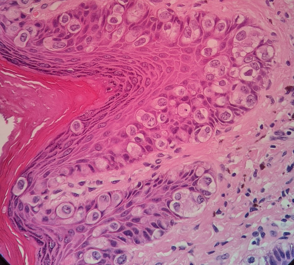
This is an example of squamous cell carcinoma of the cervix:
📍”Tadpole” abnormally shaped cell
📍Atypical keratinizing (pink) cells
📍Necrotic diathesis- granular debris
Screening and HPV vaccines help prevent these cancers! #NationalCancerPreventionMonth#CytoPath#GYNPath
CLASSIC "tissue culture" appearance of a benign mesothelial sheet, inadvertently sampled in a mediastinal FNA. The significant nuclear pleomorphism is not uncommon and such fragments should not be overcalled as "atypical".
Convencional PAP smears.
Adenocarcinoma in situ
Crowded groups of Endocervical cells, with pseudoestratificación.
Elongated nucleous
Clumpping chromatin
Mitosis
Rosette arquitecture
Feathering
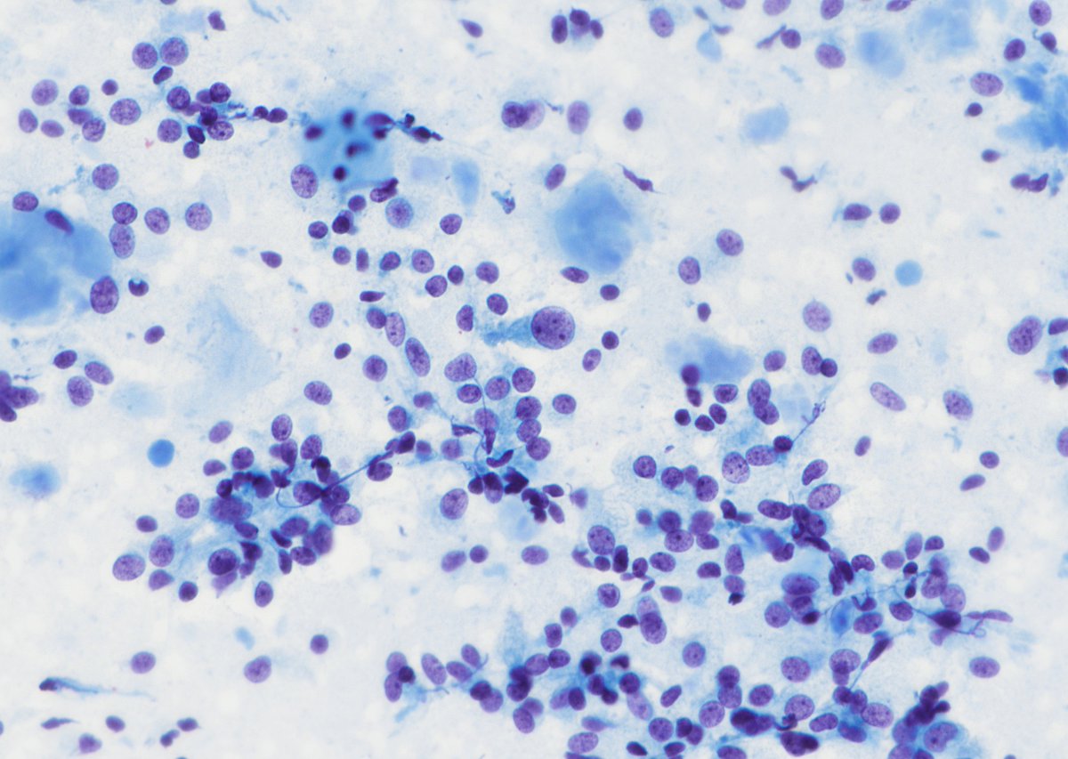
Metastatic Merkel cell carcinoma in the CSF – pt w/ a known h/o the disease and suspicion for leptomeningeal carcinomatosis by imaging. Cytology shows loosely cohesive clusters of round cells w/ high N:C ratio and characteristic finely stippled, powdery neuroendocrine chromatin.
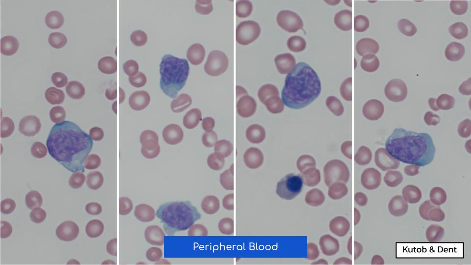
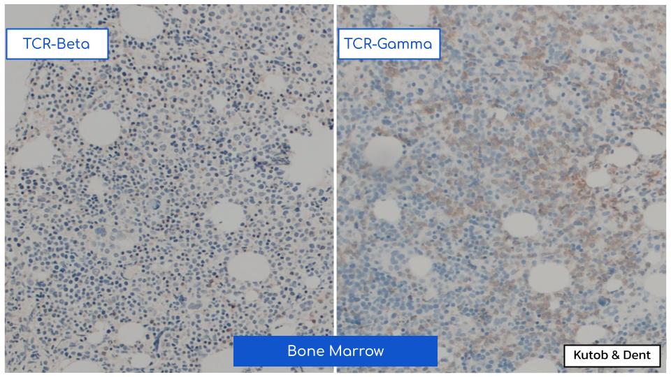
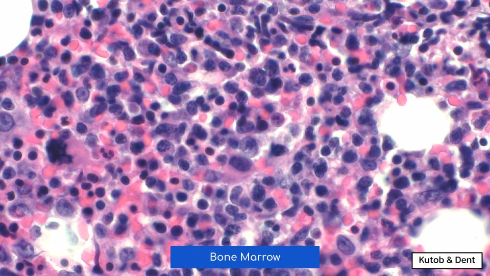
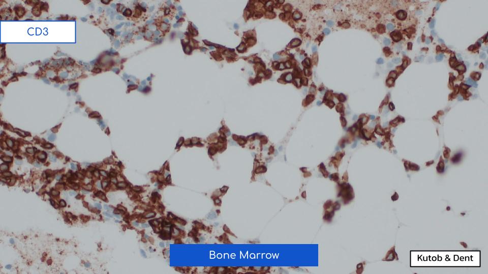
EMORY CASE OF THE WEEK #61:
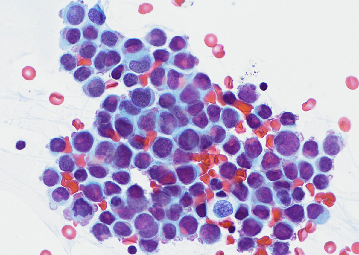
55♂️ w/ weight loss, syncope, thrombocytopenia, & suspected myelodysplastic syndrome.
Peripheral smear & bone marrow biopsy show the following.
Contributors: Leila Kutob & Alex Dent, MD (@isitcancer)
#hemepath#pathresidents#Emorypathres#EmoryCoTW