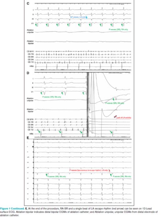
Dual-site atrial lead configuration restores intra-atrial synchrony in sinus node isolation due to intra-atrial conduction block after extensive atrial ablation
https://t.co/1fYginKXCO
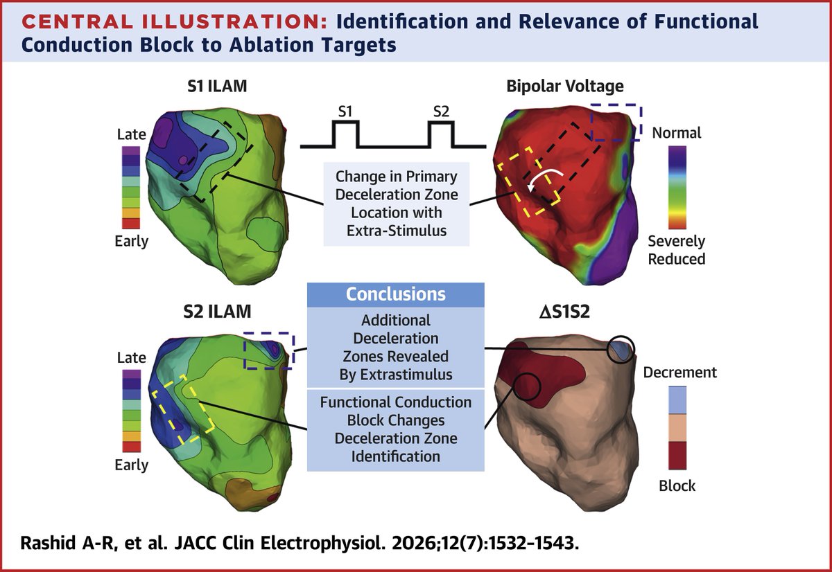
Extrastimulus affects DZ location and can reveal hidden DZs, while functional conduction block can mask DZs seen on S1 maps. ΔS1S2 maps demonstrate a visual representation of assimilation of the information within S1 and S2 maps. https://t.co/w9USuWXoc6 #JACCCEP
Early-onset DCM with NSVT in a 16-year-old highlights the value of integrating echo, CMR and genetic testing in young patients with unexplained cardiomyopathy. A novel CRYAB variant may carry important diagnostic and arrhythmic implications. https://t.co/I6hRri15Qi
#EHJCaseReports @Phiso_de@TJ_Yeo@aayshacader@girishviswa@SukritiBanthiya@O_Azizy_MD@saramoscatelli7@EHJCREiC #CardioX
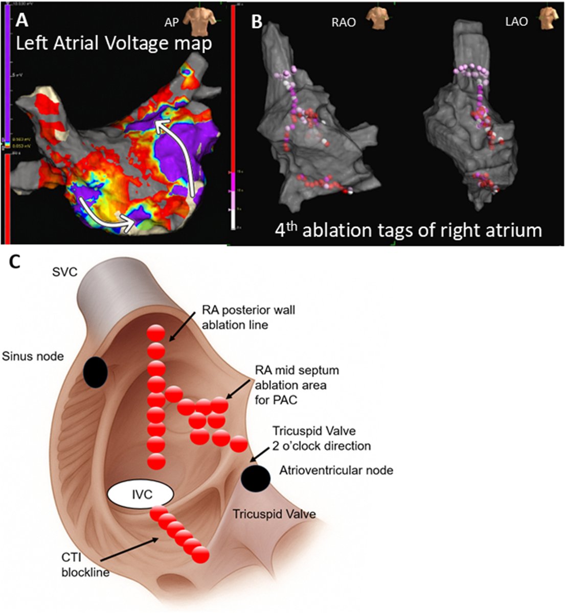
The most challenging Atach.( focal) I’ve encountered in my carrier! 😳; 7 yrs child with incessant tachy & TCM( EF:10%)! failed in OH; RA SOO around CT area but deep and repeated recurrence!never had SR for a few yrs. & mis- interpreted as inapp. ST🤷♂️!#Epeeps
This is exactly why we do #EP study in asymptomatic pre-excited patients. To risk stratify accessory pathways!
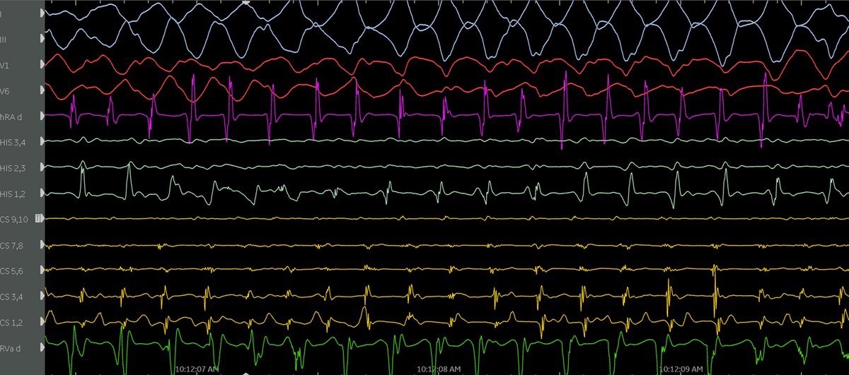
Young patient with manifest posteroseptal AP. During EP study, pre-excited sinus rhythm degenerated before our eyes:
Sinus-->AFib-->AFL 1:1--> PMVT/VF --> shocked
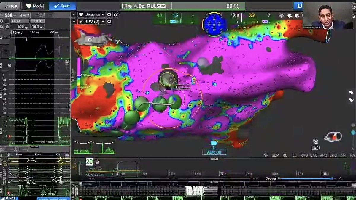
Persistent AF ablation in 2023 (PVI-Carto )
Late recurrence in 2026.
Re-do with Columbus.
Easy-star catheter in LSPV. Large potential seen.
Abl catheter in LAA.
SR and then pacing from CS.
Is LPSV reconnected?
Diagnosis and Treatment of Reentrant Atrial Tachycardia at the Superior Vena Cava-right Atrium Junction using a Dual-Energy Lattice-tip Catheter
https://t.co/fZ9IrIQKfl
Precise localization of the ventricular insertion of a bystander concealed nodoventricular pathway in east–slow atrioventricular nodal reentrant tachycardia #OpenAccess@Koichi16423232
https://t.co/eeCqZmxlmq
A multidimensional mapping array for assessment of myocardial activation using the electrotomographic mapping concept #OpenAccess@kasundesilva_
https://t.co/06AdZhnwnW
Ripple mapping demonstrates putative signals identifying the right inferior nodal extension to the lower nodal bundle during AVNRT #OpenAccess@balrik
https://t.co/GjSzHTo67i
Wide-complex tachycardia can be difficult to classify, but the limb lead algorithm provides a quick, stepwise approach using only the limb leads.
How it works:
Step 1: Is there a monophasic R wave in lead aVR?
If yes, the rhythm is ventricular tachycardia (VT).
Step 2: If not, are leads I, II, and III all predominantly negative?
If yes, this strongly favors VT.
Step 3: If neither criterion is present, examine the inferior leads (II, III, aVF). If they are all monophasic positive or all monophasic negative, check the remaining limb leads (I, aVR, aVL) for opposite polarity. If this pattern is present, the diagnosis is VT.
🟥 If these criteria are not met, the algorithm does not classify the rhythm. Additional ECG criteria and clinical assessment are required to distinguish VT from supraventricular tachycardia (SVT) with aberrant conduction.
Take-home message:
The limb lead algorithm is a practical bedside tool that can rapidly identify many cases of VT using only limb leads. However, it is not designed to diagnose every wide-complex tachycardia, so an indeterminate result should prompt a comprehensive 12-lead ECG analysis rather than exclusion of VT.
Reference: Zipes DP, Jalife J. Cardiac Electrophysiology: From Cell to Bedside.
Always amazing listening to @PrashSanders
This time talking on Non-PV isolation in Persistent Afib. Thanks from Indian Heart Rhythm Society
#afib#epeeps
A 12-lead ECG can do more than diagnose ventricular tachycardia (VT). It can also help predict where the arrhythmia exits the ventricle, guiding catheter ablation before the procedure begins.
A practical approach:
Start with the limb leads to estimate the VT exit site:
🟨 Septal
⬜ Superior
🟩 Inferior
🟥 Lateral
Next, examine the precordial leads (V1–V6) to determine whether the exit is:
🟪 Basal
🟧 Mid-ventricular
🟦 Apical
Why this matters:
- The initial QRS vector during VT points away from the exit site.
- QRS morphology helps narrow the region where the reentry circuit exits the myocardium.
- Reviewing the clinical VT ECG before ablation improves procedural planning and mapping efficiency.
Prior myocardial infarction also influences VT morphology:
- Anterior infarction (LAD territory) produces characteristic anterior scar-related VT patterns.
- Lateral infarction (LCx territory) results in distinct lateral exit morphologies.
- Inferior infarction (RCA territory) generates different inferior scar-related VT patterns.
If the clinical VT ECG is unavailable, ICD electrograms and pace mapping can help confirm the VT mechanism and estimate the catheter's proximity to the exit site during the procedure.
Reference: Zipes DP, Jalife J. Cardiac Electrophysiology: From Cell to Bedside, Chapter 86.