Localizing Myocardial Infarction via ECG Leads
A complete coronary blockage progresses through: Ischemia → Injury → Infarction.
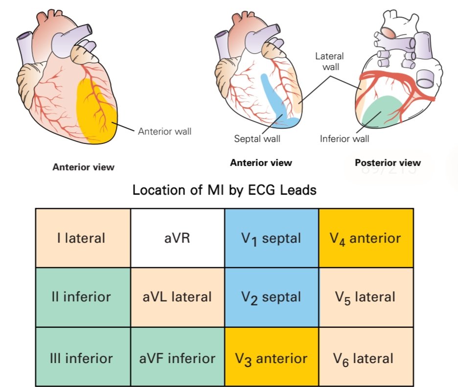
MI location is key for guiding treatment and predicting complications. ECG leads help pinpoint the affected heart wall:
▶️ Anterior MI: V3, V4
▶️ Septal MI: V1, V2
▶️ Lateral MI: I, aVL, V5, V6
▶️ Inferior MI: II, III, aVF
▶️ Posterior MI: reciprocal changes in V1–V3
Clinical Tip: An MI may not be limited to a single region.
Example: Changes in V3, V4 (anterior) + I, aVL, V5, V6 (lateral) indicate an anterolateral MI.
Recognizing patterns across leads is critical for accurate diagnosis and timely intervention.
dok aku sudah selesai iship dan berencana untuk apply PTT buat dapetin rekom PPDS, tapi galau apakah ini langkah yg tepat untuk langsung ambil PTT? krn katanya nanti dapet rekom bupati s.d spesialis yg dituju. Tapi takut untuk daftar PPDS surat rekom itu juga blm cukup (cont..)