🧬 Spatial biology is moving beyond the transcriptome.
A timely JCI Insight review maps the rapidly expanding landscape of spatial multiomics—technologies designed to measure not only where genes are expressed, but also where genomic alterations, epigenetic states, proteins, metabolites, and functional phenotypes occur within intact tissues.
Spatial transcriptomics has transformed biomedical research, but RNA alone provides an incomplete picture. mRNA abundance may correlate poorly with protein levels or epigenetic states and cannot directly capture metabolic activity or small-molecule distributions.
The next generation therefore asks:
Can we reconstruct multiple layers of biology while preserving tissue architecture?
The review organizes this rapidly evolving field into several major dimensions:
🧬 Spatial genomics
Methods such as Slide-DNA-seq can map genomic alterations directly within tissues, allowing investigators to connect tumor clones with their spatial niches and distinguish genetically driven from microenvironment-dependent transcriptional programs.
🔐 Spatial epigenomics
Spatial-ATAC-seq and spatial-CUT&Tag reveal chromatin accessibility and histone modifications in situ. Newer approaches can simultaneously capture epigenome + transcriptome + proteins, moving toward genuine spatial tri-omics.
🧪 Spatial proteomics
Sequencing-based, imaging-based and mass-spectrometry platforms provide information that RNA cannot: protein abundance, localization, posttranslational modifications and potentially protein-protein interactions within disease niches.
⚡ Spatial metabolomics
MALDI-MSI, DESI-MSI and label-free optical approaches map metabolites, lipids, redox states and even drug distributions—providing a more direct readout of cellular function and pharmacology.
One particularly exciting frontier is spatial functional genomics: combining CRISPR perturbation with spatial readouts to determine how specific genetic changes reorganize neighboring cells and tissue ecosystems. Platforms including Perturb-map, Perturb-Multi and PERTURB-CAST are beginning to turn spatial atlases from descriptive maps into causal experiments.
And AI may dramatically accelerate clinical translation.
Instead of experimentally measuring every molecular layer, emerging models attempt to infer spatial molecular states directly from routine H&E histopathology, potentially creating scalable “virtual spatial profiling.”
The field still faces major challenges: cost, resolution-versus-coverage tradeoffs, cell segmentation, cross-platform normalization, multimodal alignment and standardized benchmarking.
But the trajectory is clear:
Spatial transcriptomics → spatial multiomics → spatial functional biology → predictive tissue models.
The ultimate goal is no longer simply to build increasingly detailed cell atlases.
It is to understand how genome, epigenome, transcriptome, proteome and metabolome interact in space to create disease—and therapeutic response.
📄 Mao X, Chen Z, Hwang EJ, et al. Spatial multiomics in biomedical research: advances beyond transcriptomics.
JCI Insight. 2026;11(17):e206951
🔗 DOI: 10.1172/jci.insight.206951
#SpatialBiology #SpatialOmics #Multiomics #SpatialTranscriptomics #Proteomics #Metabolomics #Epigenomics #SingleCell #PrecisionMedicine #AI
We updated our Susztaklabbiobank website https://t.co/IHMd3V0EW7
For almost two decades our laboratory has collected and profiled human kidney tissue, paired it with mouse and rat models and with the genetics of millions of people, and built one of the most complete maps of the human kidney that exists. This site is where we share it: genome-wide association results, expression, methylation, protein and open-chromatin maps, single-cell and single-nucleus atlases, spatial transcriptomes and animal-model resources, each in an interactive browser you can query today. Please send your suggestions how we could improve our site. Let's cure kidney disease together !
Check out our recent review @JCI_insight on spatial multiomics technologies and their applications! The field is evolving so fast😍
Spatial multiomics in biomedical research: advances beyond transcriptomics https://t.co/v4zBOBTZv7
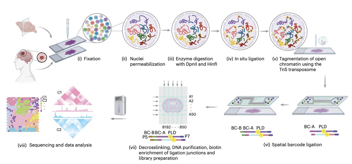
Excited to share Spatial-ATAC-Hi-C @naturemethods, spatial profiling of 3D genome organization + chromatin accessibility in tissue. Excitingly, it detects CNVs and SVs in tumors and reveals spatial heterogeneity.
Great collaboration with @RongFan8.
https://t.co/XO0FhoZepO
Excited to share that Spatial Hi-C-RNA is now out in @CellCellPress! It brings the 3D genome into spatial multi-omics by co-mapping chromatin architecture and gene expression in the same tissue section. Congrats to the whole team! https://t.co/ov9hQkKNOA
Excited to share our new paper out in @NatureComms !
We introduce ArchVelo, a new computational framework for RNA velocity and trajectory inference from single-cell multi-omic (scATAC+RNA-seq) data:
https://t.co/YCjbHhhUQd
Plasma proteins can predict metabolic liver disease (MASLD) up to 16 years ahead of its clinical appearance.
One study after another showing the forecasting value of proteomics
https://t.co/mqgQ1INuCB
Here is the surprise. The textbook says that once a cell commits, its fate is set. We found that this is not the whole story. Proximal tubule cells — already carrying a mature tubular identity — can revise their fate and become parietal epithelial cells.
Two independent trajectory methods, RNA velocity and random-walk analysis, pointed the same way: from proximal tubule toward parietal cell. Staining caught the act — cells with parietal shape still expressing ACE2, a proximal tubule marker. Only in fetal, never adult, kidney.
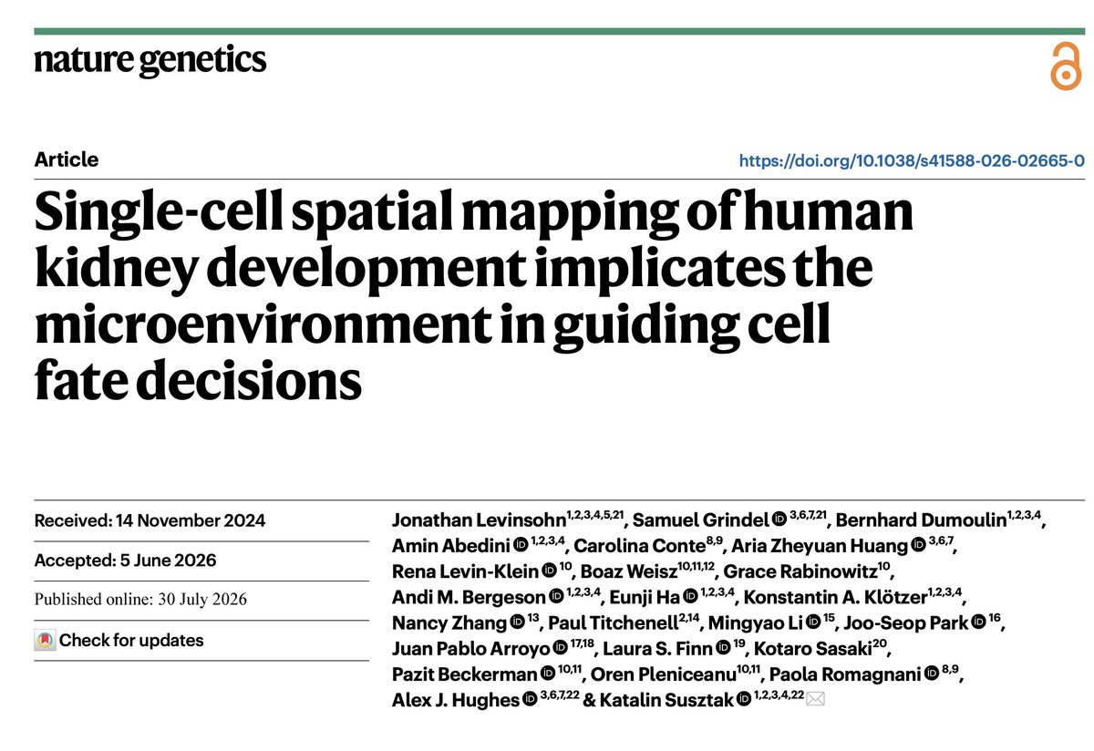
🔥🎆Our newest paper is out !!! @NatureGenet How does the kidney build its filtering units, each from the same small pool of stem cells? Jon Levisohn, Alex Hughes lab mapped human kidney development cell by cell, in space, and found it is far more flexible than the textbook teaches. https://t.co/58gdC3ZzpU
Excited to share Pan-human Azimuth, an NIH @_hubmap reference for single-cell data https://t.co/PzQ1Z7Sccl
We invested deeply in human curation of training data, prioritized diversity+breadth+extensive QC, and uniformly labeled cells across 23 tissues in a single hierarchy (1/)
Excited to share that our Universal Cell Embedding (UCE) paper is published in @Nature ! Single-cell RNA sequencing data gives us an unprecedented look into the diversity of cell biology, but analysis has often been limited to the specific dataset or atlas that was collected.
https://t.co/FmH1WDd23F
Totally over the moon 🤩 , our paper on charting human cellular senescence in aging and disease is on the cover!! It highlights the collective efforts and the first wave of publications from NIH @sennetresearch consortium to map senescent cell states, heterogeneity and niches!!
🤩 spatial panoramic in vivo CRISPR screen via Perturb-DBiT 🤩 Excited to share our collaborative work with @sidichen lab and many outstanding collaborators, now published in Nature Biotechnology!
https://t.co/swMT8pJajh
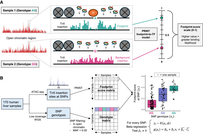
Characterization of non-coding variants associated with transcription-factor binding through ATAC-seq-defined footprint QTLs in liver
https://t.co/ncDF7vvO3l
Highlight from @AJHGNews (@GeneticsSociety)
@CHOP_Research@cambupenn