How does a cardiac muscle cell contract and relax?
Every heartbeat depends on a tightly coordinated process called excitation-contraction coupling, which links the electrical action potential to mechanical contraction of the myocardium.
The sequence is straightforward:
◻️An action potential travels along the sarcolemma and deep into the cell through the T-tubules.
◻️Depolarization opens L-type calcium channels, allowing a small amount of calcium to enter the cell.
◻️ This incoming calcium activates ryanodine receptors (RyR) on the sarcoplasmic reticulum (SR), triggering a much larger release of calcium into the cytoplasm. This process is known as calcium-induced calcium release.
◻️ The rise in intracellular calcium allows calcium to bind troponin C, exposing binding sites on actin so that myosin heads can attach and generate force using ATP.
◻️ During systole, repeated actin-myosin cross-bridge cycling pulls the thin filaments toward the M-line, shortening the sarcomere and producing myocardial contraction.
Relaxation requires rapid removal of calcium from the cytoplasm:
◼️ SERCA (SR Ca²⁺-ATPase) pumps most calcium back into the sarcoplasmic reticulum. Its activity is regulated by phospholamban (PLB).
◼️The Na⁺/Ca²⁺ exchanger (NCX) removes calcium from the cell by exchanging one calcium ion for three sodium ions.
◼️A smaller amount of calcium is extruded by the sarcolemmal Ca²⁺-ATPase or taken up by mitochondria through the mitochondrial calcium uniporter.
Structurally, the contractile apparatus consists of:
🟥Thin filaments: Mainly actin
🟩Thick filaments: Myosin
⬜Z-lines: Boundaries of each sarcomere
🟪M-line: Central region where myosin filaments are anchored
🟨Titin: A giant elastic protein that stabilizes the sarcomere and contributes to passive myocardial stiffness and recoil.
The coordinated movement of calcium and ATP-driven cross-bridge cycling allows the heart to contract forcefully during systole and relax efficiently during diastole, enabling continuous pumping throughout life.
Reference: Braunwald's Heart Disease: A Textbook of Cardiovascular Medicine, Figure 46.1.
How long should anticoagulants be withheld before elective surgery?
◻️Apixaban: 1 day (low/moderate bleeding risk), 2 days (high risk)
◻️Rivaroxaban: 1 day (low/moderate), 2 days (high)
◻️Edoxaban: 1 day (low/moderate), 2 days (high)
◻️Dabigatran: 1-4 days depending on bleeding risk & renal function
◻️ Fondaparinux: 36-42 hours
◻️ LMWH: 12 h (prophylactic), 24 h (therapeutic)
◻️ UFH: IV 4-6 h; SC 12–24 h
◻️ Warfarin: Stop 5 days before surgery; confirm INR before the procedure.
Adjust timing for renal impairment, neuraxial anesthesia, and individual patient risk. Bridging is reserved for selected high-thrombotic-risk patients.
Reference: 2024 AHA/ACC Guideline for Perioperative Cardiovascular Management for Noncardiac Surgery, Table 13.
A new review summarizes advances in the treatment of multiple sclerosis, with emphasis on effective disease-modifying therapies for relapsing disease and the need for better treatments to prevent progression and rebuild myelin.
Read the Review Article “Advances in Multiple Sclerosis” by Stephen L. Hauser, MD: https://t.co/McH5l7urUG
NEJM subscribers: Try AI Companion to surface key details from this article instantly.
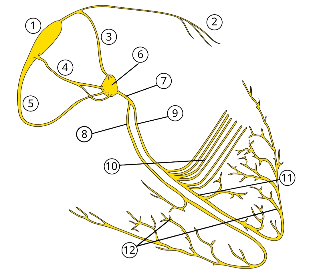
Electrical conduction system of the heart:
1. Sinoatrial (SA) Node: Often called the heart's natural pacemaker, it initiates the electrical impulse.
2. Bachmann's Bundle (Interatrial Tract): Conducts the impulse from the right atrium to the left atrium.
3. Anterior Internodal Tract: One of three pathways connecting the SA node to the AV node.
4. Middle Internodal Tract (Wenckebach's pathway): The second internodal pathway.
5. Posterior Internodal Tract (Thorel's pathway): The third internodal pathway.
6. Atrioventricular (AV) Node: Acts as a gatekeeper, briefly delaying the signal to allow the ventricles to fill with blood.
7. Bundle of His (Atrioventricular Bundle): The bridge that carries the signal from the atria into the ventricles.
8. Right Bundle Branch: Carries the electrical impulse to the right ventricle.
9. Left Bundle Branch: Carries the electrical impulse to the left ventricle.
10. Left Anterior Fascicle: A branch of the left bundle that stimulates the upper/front part of the left ventricle.
11. Left Posterior Fascicle: A branch of the left bundle that stimulates the lower/back part of the left ventricle.
12. Purkinje Fibers: The final network of fibers that spread the impulse throughout the ventricular muscle, causing contraction.
》Short debate questions:
“Are the classical anterior, middle, and posterior internodal tracts real structures, or just functional pathways in atrial myocardium?”
“If discrete internodal tracts are not clearly proven anatomically, how does the impulse travel so quickly from the Sinoatrial Node to the Atrioventricular Node?”
🩸 Cancer-Associated VTE | Extended Apixaban – Bleeding Predictors (API-CAT) 🩸
📌 Lancet Haematology 2026 – Post-hoc API-CAT analysis
�� Who bleeds more on extended anticoagulation (>6 months)?
⚠️ Independent predictors of clinically relevant bleeding:
👴 Age ≥75 years
🩸 Anemia (Hb <10 g/dL) and/or thrombocytopenia (<100,000/µL)
🫁 Index event = Pulmonary embolism
👨 Male sex
📊 Key points:
✅ Same predictors across cancer sites
✅ No interaction with apixaban dose (2.5 vs 5 mg BID)
✅ Cumulative bleeding risk ↑ as predictors accumulate
❌ Cancer site & metastatic status NOT predictive after 6 months
🧠 Clinical pearl:
After 6 months of CAT treatment, bleeding risk is driven more by patient biology than tumor factors → personalize duration & intensity of anticoagulation.
📚 Source (high-quality, peer-reviewed):
The Lancet Haematology, Jan 2026 – API-CAT investigators
https://t.co/pMXakxTwrO
#Hematology #Thrombosis #CancerAssociatedThrombosis #DOACs #Apixaban #PatientSafety #EvidenceBasedMedicine
Drugs Causing Acute Interstitial Nephritis (AIN)
Mnemonic: RONALDO
R – Rifampicin
O – Omeprazole
N – NSAIDs
A – Allopurinol, Antivirals
L – Beta-lactam antibiotics
D – Diuretics
O – Ofloxacin
A school-age child, previously fluid, presented vesiculobic lesions to both hands, with a 6-day evolution. According to the family members, the injuries started as small vessels and gradually spread in a centrifugal way, forming concentric rings in a spiral pattern.
The injuries were asymptomatic, without systemic symptoms, and there was no recent history of travel, environmental exposure, use of topical agents or contact with sick animals or individuals. There were no accommodation in other parts of the body. What is the probable diagnosis?
Your immune system doesn't only defend, it remembers
This diagram maps how the body’s innate and adaptive immune systems work together to identify, attack, and remember pathogens. It shows how different immune cells, antibodies, and signaling molecules coordinate to protect against infection while maintaining balance.
1️⃣ Innate immunity: the immediate response
The innate system acts within minutes, providing broad defense through physical, chemical, and cellular barriers.
🟢 Example: Skin, mucus, and stomach acid block pathogens, while macrophages, neutrophils, and natural killer cells identify and destroy invaders using pattern-recognition receptors.
🟢 Example: The complement cascade amplifies inflammation and flags pathogens for destruction by immune cells.
2️⃣ Adaptive immunity: the targeted response
The adaptive system learns to recognize specific antigens and produces lasting protection through specialized B and T cells.
🟢 Example: B cells release antibodies (IgM, IgG, IgA, IgE, IgD) that neutralize toxins and tag microbes for clearance.
🟢 Example: T cells coordinate and execute defense—helper T cells activate macrophages and B cells, while cytotoxic T cells trigger apoptosis in infected cells.
3️⃣ Antibody specialization and immune memory
Each antibody class has a unique role in defense and long-term immunity.
🟢 Example: IgM is the first antibody made in infection, IgG provides long-term protection and crosses the placenta, IgA guards mucosal surfaces, and IgE mediates allergic reactions.
🟢 Example: Memory B and T cells remain after infection or vaccination, allowing faster and stronger immune responses upon re-exposure.
4️⃣ Active and passive protection
Immunity can be acquired through natural infection, vaccination, or temporary antibody transfer.
🟢 Example: maternal antibodies passed through breast milk provide short-term passive defense.
The immune system’s strength lies in its coordination—an instant, non-specific response that buys time for a targeted, adaptive defense that remembers what it has seen.
Violin-string adhesions between the liver capsule and abdominal wall are classically associated with which pathogen?
A) Escherichia coli
B) Neisseria gonorrhoeae
C) Hepatitis B virus
D) Entamoeba histolytica
🔥 The Heart Failure Meds Mnemonic Every Medico Should Know
💊 First-line HF drugs → BAD ANL
📍 B — Beta-blockers
📍 A — ACE inhibitors
📍 D — Diuretics
📍 A — ARNI (Sacubitril/valsartan)
📍 N — Nitrates + Hydralazine
📍 L — Loop diuretics for congestion
💬 Team ACEI or Team ARNI❓ Drop your vote 👇
● NOACs Overall Clinical Takeaways:
▪︎ Renal function is key for drug selection and dosing — Dabigatran has highest renal clearance.
▪︎ Apixaban is the most kidney-friendly.
▪︎ Rivaroxaban must be taken with food at higher doses.
▪︎ Dabigatran is unique: prodrug, dialysable, and acid-sensitive.
▪︎ CYP interactions most relevant for Apixaban and Rivaroxaban.
⚡ Quick Key Reminders
🧠 Only prodrug: Dabigatran
💊 Only thrombin inhibitor: Dabigatran
💧 Dialysable: Only Dabigatran
⚠️ Most CYP interactions: Apixaban, Rivaroxaban
⚕️ Safest in renal impairment: Apixaban
🍽️ Food required: Rivaroxaban
🔄 Balanced clearance: Edoxaban
Table from EHRA practical guide
What else would you like to add? 🤔
NOACs in treatment of DVT/PE
Apixaban and Rivaroxaban start as oral monotherapy (no need for initial heparin).
Dabigatran and Edoxaban require 5–10 days of parenteral anticoagulation before starting >>>
(this is sequential rather than overlapping bridging). Why so?!
Table from EHRA practical guide