Issue 13 is complete!
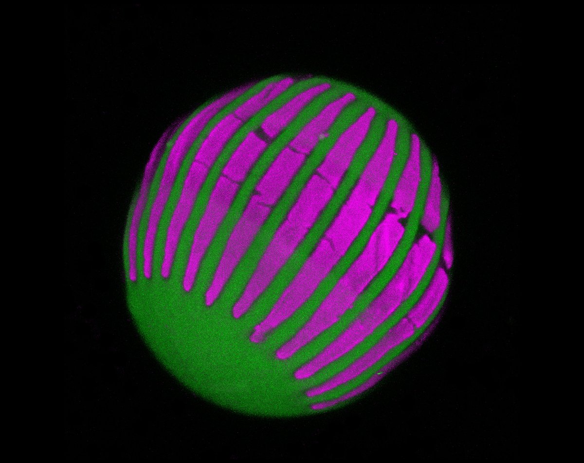
On the cover: zebrafish cardiac ECM labelled by Tg(ubb:ssNcan-GFP) & surface rendered in 3D using Imaris. Colors mark distinct cardiac regions: ventricle (purple), atrioventricular canal (yellow) & atrium (green). See Gentile et al.
https://t.co/PvWndJ95Bv
📣 NEW IMARIS 10.2 📣
The newest Imaris 3D image analysis software brings faster data rendering for all users. In addition, all Imaris 10.2 functionality, including AI segmentation is now faster on Apple M3 processors.
Take a 10 day Free Trial here ⤵️
https://t.co/MDwMtyRM3e
📣 NEW IMARIS 10.2 📣
The newest Imaris 3D image analysis software brings faster data rendering for all users. In addition, all Imaris 10.2 functionality, including AI segmentation is now faster on Apple M3 processors.
Take a 10 day Free Trial here ⤵️
https://t.co/lUgdNvG3j5
I'm giving a webinar next month! Want to learn how to do 3D high-resolution brain microscopy analysis?
Register using the link below; it is free! ���🔬🧠 https://t.co/SnEBEy4TpN
@ImarisSoftware
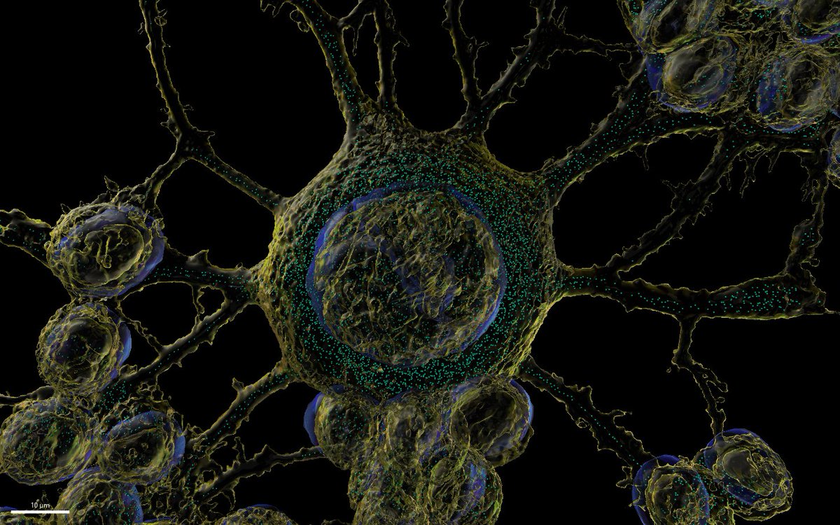
Congrats @CHIRI_curtin image of the month winner Dr Gae Ellison!
Iron (ferroorange) in neuroblastoma cells, rendered using spots and surfaces in @ImarisSoftware
Captured using @AndorTechnology Dragonfly using @NikonInst 60x lens
Our end-to-end workflow for 3D pathology is now published in @NatureProtocols!
This includes all the steps to go from archived pathology tissues to 3D H&E-like datasets, with an emphasis on quality control for large studies.
Full text at: https://t.co/MfPzUCe3aJ
2 More movies (that I forgot to post🤡).
1st is human embryo skeleton; 2nd is inner and middle ear . Segmented with @syGlassVR@ImarisSoftware and rendering with @BlenderStudio_