Our latest work on the HOPS tethering complex is now out in MBoC special issue 'Cell Biology of the Lysosome'! We show pathways affected by depletion of HOPS, including impaired endosomal maturation and recycling, but amphisomes still form?! Read more: https://t.co/RGxSVCRLKt
We recently published a JoVE article and video on how we do on-section CLEM in our lab! It contains useful insights, tips and tricks for cryosectioning, labeling on sections and doing LM-EM correlation. Below is just a snippet, find the full protocol at: https://t.co/I8kJOiWSD0
Imaging lysosomes in 3D cultured cells on a lattice light sheet of the @AICjanelia with Chad Hobson has been amazing! We leave with answers, more questions and a healthy 62 TB of data, only possible because of the great support here. A big thank you to Chad and the AIC 😊
@JKlumperman@NalanLiv Winning this prize made me surprised, humbled and very grateful. Warm thanks to Judith and Nalan for the nomination and the Institute of Biomembranes and selection committee for granting the award.
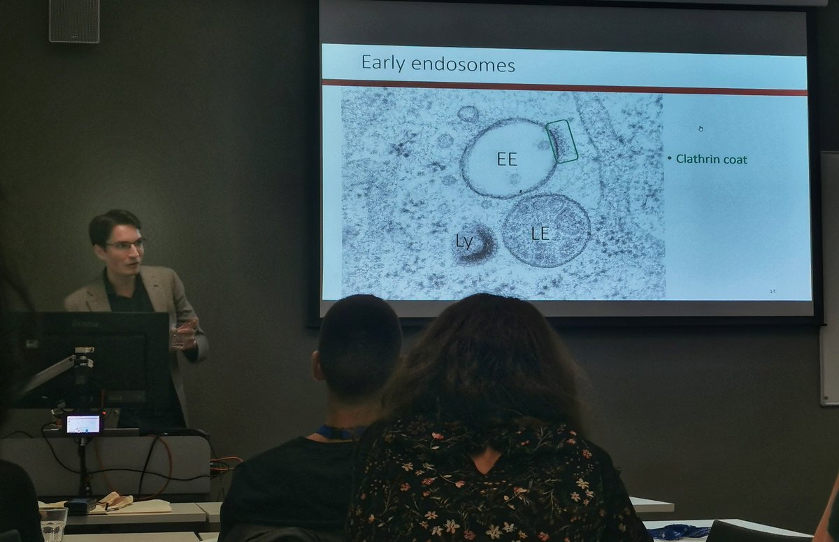
Now we host @JA_Beek, who's teaching us novel lessons with the good old tools (🔬).
Wanna check the whole endolysosomal biogenesis pathway under the electron microscope lens? 🧐
❕Stay tuned for this exciting talk 🤗
On the super-hot HOPS complex and its overall function, we'll have 👨🏫Jan van der Beek (@JA_Beek), who's a PhD in the lab of 👩🏫Judith Klumperman (🇳🇱).
#ESGLD#graduatecourse#teachers
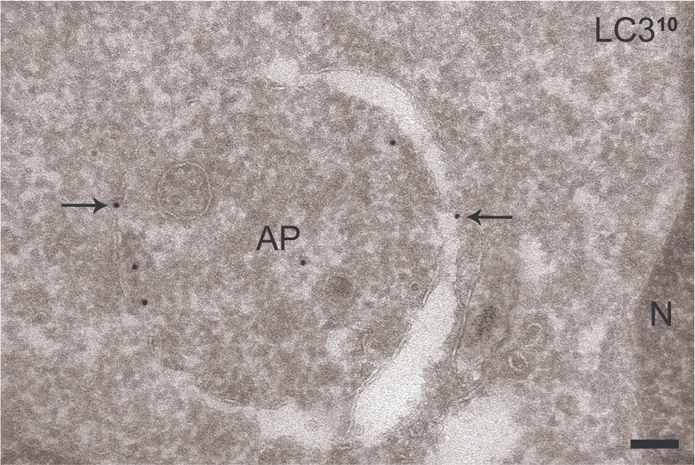
Just out, a new toolbox article from Ann De Mazière, me and others from the @JKlumperman lab! In this paper in Autophagy, we describe an optimized method for labeling LC3 in Electron Microscopy. A valuable tool for all researchers in #autophagy!
https://t.co/Ok4srzlg14
In light of all the recent developments, probably more people need to see this.
A fight against hopelessness.
We CAN and SHOULD continue our efforts to reduce emissions.
https://t.co/6ya44qX3xp
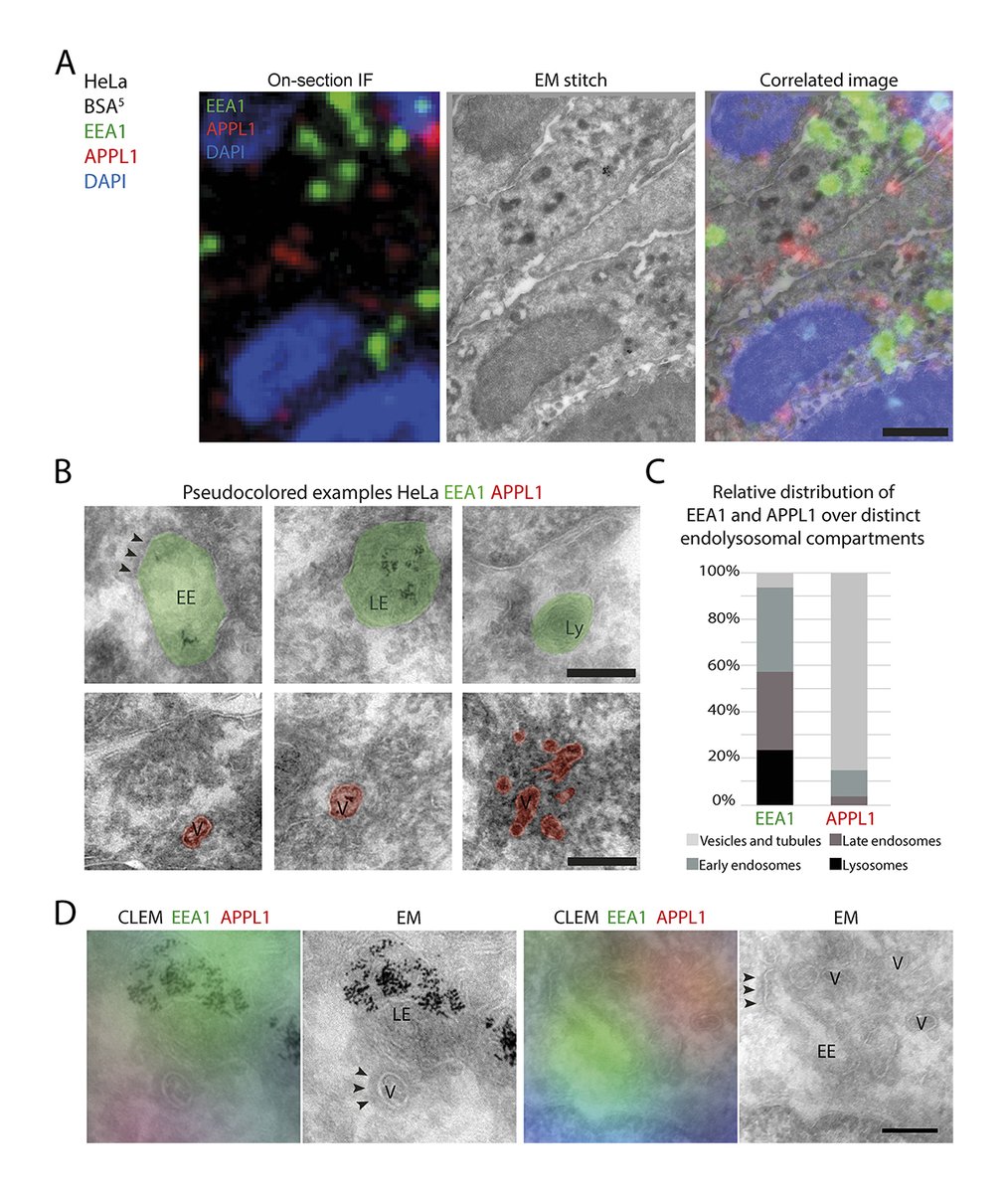
I'm very happy to share our latest work in which we use CLEM to study key endo-lysosomal proteins. It's now out in JCB and includes a new dataset on PI(3)P. It localizes together with EEA1 on late endosomes, indicating a broader ultrastructural distribution than commonly thought.
A quantitative CLEM method by @JA_Beek, de Heus, @NalanLiv & @JKlumperman determines the ultrastructural distribution of Rab5, Rab7, EEA1, APPL1 & PI(3)P, revealing distinct endosomal subpopulations & an unexpected localization of EEA1 to late #endosomes https://t.co/FnMmsUcic9
We’ve also gotten a facelift! 💁♀️ Browse all the related data for these papers and more - AND - volume renderings are now available for most segmentations. Check it out on https://t.co/2sHzCyO5zC !
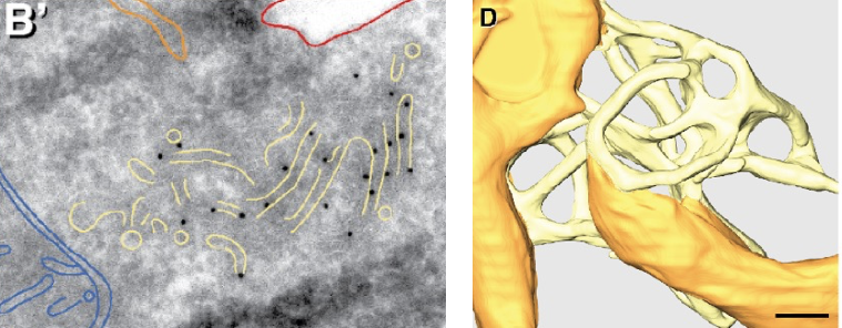
Proud to be part of a fantastic collaboration now published in Science: DOI: 10.1126/science.abh2474. Beautiful example of how complementary cryo- and immuno-EM methods converge in revealing a new ER subdomain involved in IRE1alpha signalling in stressed human cells. Part 1/2
The 2021 issue of Current Opinion in Cell Biology on Membrane Trafficking is out! Thanks to all contributing authors and to @TPucadyill for the fantastic editorial collaboration. https://t.co/JxmhHB5BSf
I'm very happy to share our latest work! We adapted our CLEM protocol to correlate large tilesets of sensitive IF and EM images. We use it to reveal the morphology behind Rab5, EEA1, APPL1 and Rab7 organelles. It's now out on BioRXiV! https://t.co/rFu9PZfdVv