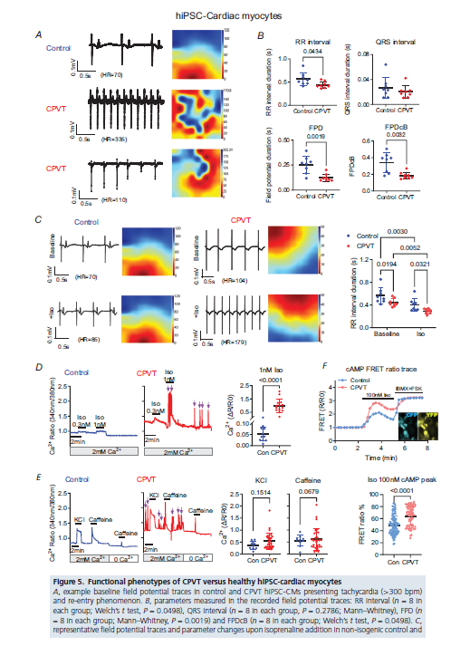
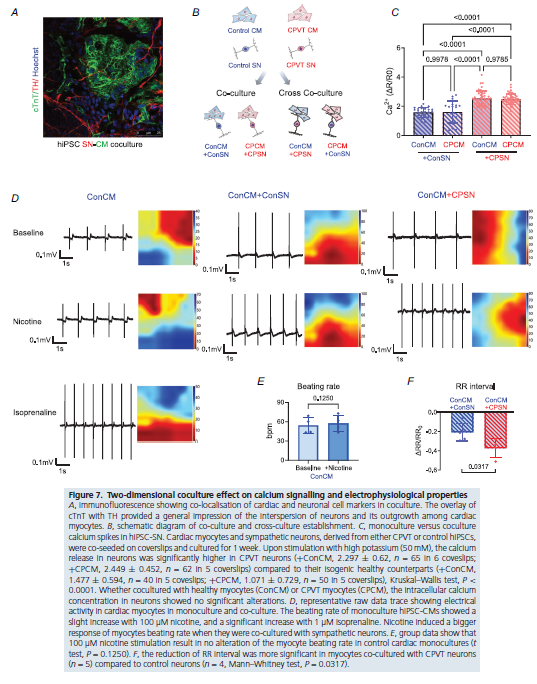
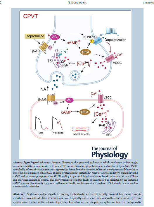
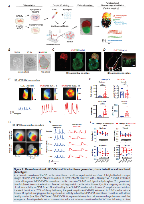
A new study shows that inherited arrhythmias like CPVT are not driven by cardiomyocytes (CM) alone.
Using hiPSC-derived neuro-cardiac models, the team found dysfunctional sympathetic neurons can trigger #arrhythmia in otherwise healthy CM.
#electrophysiology#cardiology#mapping
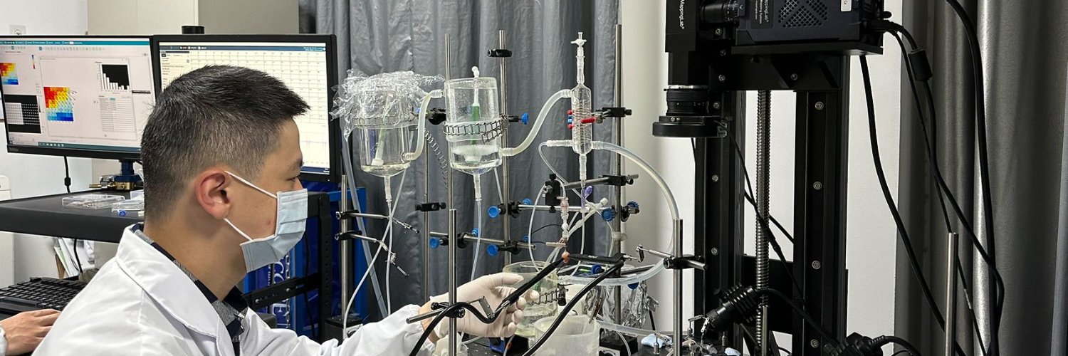
We are excited to announce the successful installation of our optical mapping system in the laboratory of Prof. Nipavan Chiamvimonvat at the University of Arizona. The system is now fully operational and provides high-resolution, synchronized mapping of cardiac AP and Ca signals.
The first gene-edited miniature pig model of LQT2 is here.
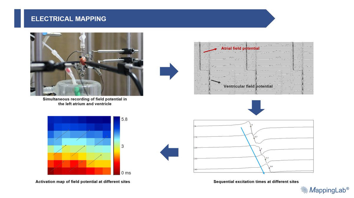
By combining ECG telemetry, electrical mapping, and optical mapping, this study delivers a truly translational view of hERG-related arrhythmias in a large-animal heart.
Short version:
https://t.co/IHscHiHlvN
Yu et al. (2025) showed that an Andr-loaded atrial patch prevents POAF by reducing inflammation, oxidative stress, and calcium imbalance. In a rat model, it lowered Afib incidence from 90% to 20% and protected heart cells, highlighting a promising targeted therapy for POAF.
Unlock the mysteries of cardiac arrhythmias with our advanced Electrical Mapping System! High precision data acquisition and MEA provide unparalleled insights into electrical activity at the tissue level. Perfect for anti-arrhythmic drug screening and toxicity testing.
Features:
◉ Precise analysis of Ca waves, transients, E–C coupling, and sarcomere length
◉ >3000 FPS CMOS imaging with 95% QE and <1 e⁻ noise
◉ All-in-one system with dual-dye recording across 400–800 nm.
◉ High-throughput Ca imaging in 10+ cells simultaneously