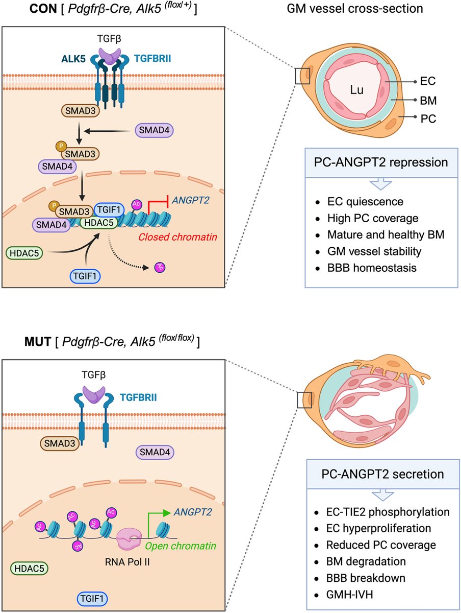
#STROKE#OriginalResearch: @juimdave et al. discover that the TGFβ pathway represses angiopoietin-2 in brain pericytes during development and identify pericyte-derived angiopoietin-2 as a target for germinal matrix hemorrhage. #AHAJournals https://t.co/NU4osZqEh2
Jui Dave, PhD, investigates potential therapeutic targets for obstructive arterial diseases, including supravalvular aortic #stenosis@GreifLab.
Congratulations @juimdave on your Career Development Award from @AHAScience. https://t.co/teEDoi798z
@YaleMed@YaleIMed
A study by @juimdave from the @GreifLab has identified the role of Notch3 in vascular development. In a laboratory setting, treatment with a pharmacological inhibitor or the genetic deletion of Notch3 has a restorative effect on the aortic wall. https://t.co/i8wDXqYNpt
@YaleMed
Postdoctoral fellow @JuiMDave aims to unravel the cellular and molecular mechanisms of cardiovascular disorders.
She is the first author of a recent @Dev_Cell study: https://t.co/9jqqYliC62
@YaleMed@YaleIMed@GreifLab@YaleCVRC
@susan_redward Most aortic diseases have defects in elastin, suggesting that elastin is absolutely integral for healthy vessel function. The interesting thing is that all of the elastin in your body is synthesized only during birth and early age. This pool will last your entire lifespan :)
@susan_redward The green is the cell nuclei (center of the cell which holds DNA). The cells are arranged in concentric rings or layers and each layer is seperated by a protein called elastin (magenta fibers). The elastin renders elasticity and mechanical support to the aorta!
@susan_redward Hope your husband is doing well now. Imagine the aorta as cylinder which helps carry the blood from heart to remaining parts of the body. You are looking at the cylinder wall cross-section. Blue stain are smooth muscle cells which majorly make up the aortic wall.