Super excited about this! Check out manuscript from my amazing postdoc Eric Mulhall and collaborators measuring single molecule #PIEZO1 dimensions in cells with nanometer precision using super-resolution fluorescence microscopy: https://t.co/RjOv8VrKOU
1/9
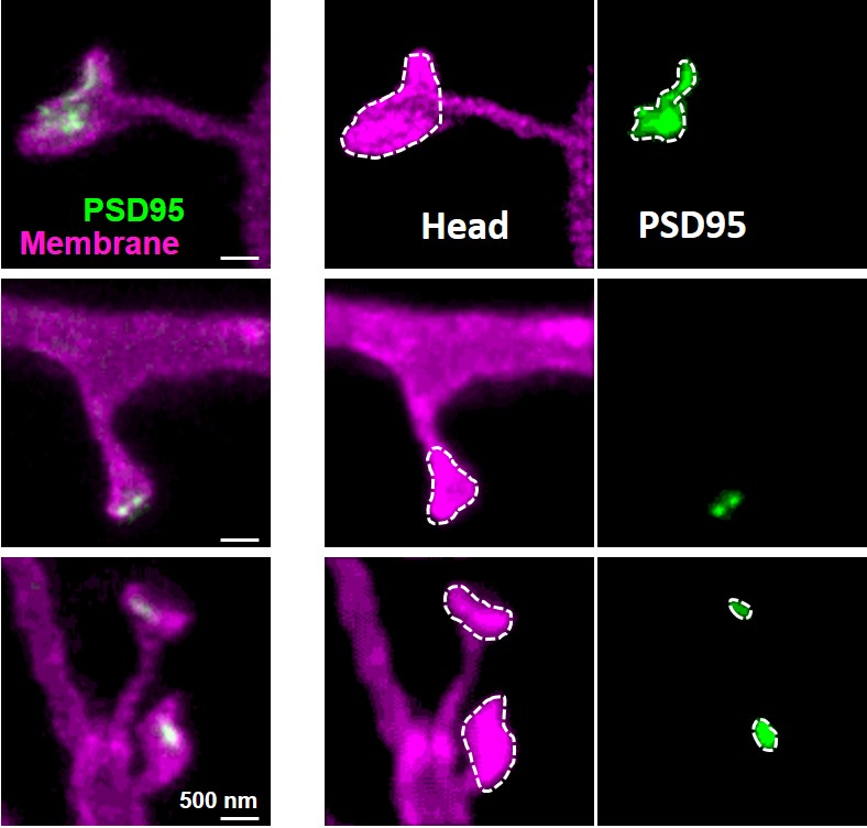
Finally out as preprint: Pre- and postsynaptic #nanostructures increase in size and complexity after #LTP induction revealed by #superresolution#STED microscopy. PhD work by Valérie Clavet-Fournier. https://t.co/LwfXwVnBSJ
Thanks @gsk_swetha for tagging me on the paper. It took a long time, but things finally fell into place. I would like to thank @AlisonBarth@KatrinWillig, and my other co-authors Matt Mosso, Joe Christian, Eunsol Park, and Waja Wegner.
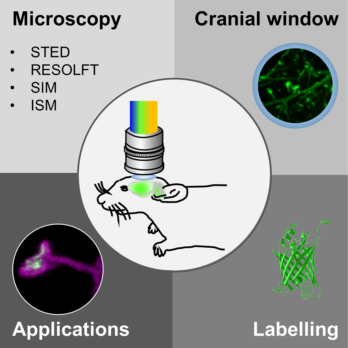
Super-resolution microscopy techniques, which offer superior spatial resolution, are now widely accessible but are rarely used in vivo. In this review, I discuss the pros and cons of potential super-resolution techniques for in vivo use to encourage a wider application.
Review article published @iScience_CP:
In vivo super-resolution of the brain – How to visualize
the hidden nanoplasticity? Thanks to @mpi_nat for support. #STED#RESOLFT#2P#SIM#ISM#super-resolution #invivo
https://t.co/iM0JOGapgw
Changes in synaptic connectivity are one of the most fundamental processes underlying the formation, storage and retrieval of memory. While 2P microscopy has opened the door to imaging of living tissue and small organisms, its spatial resolution is not sufficient ...
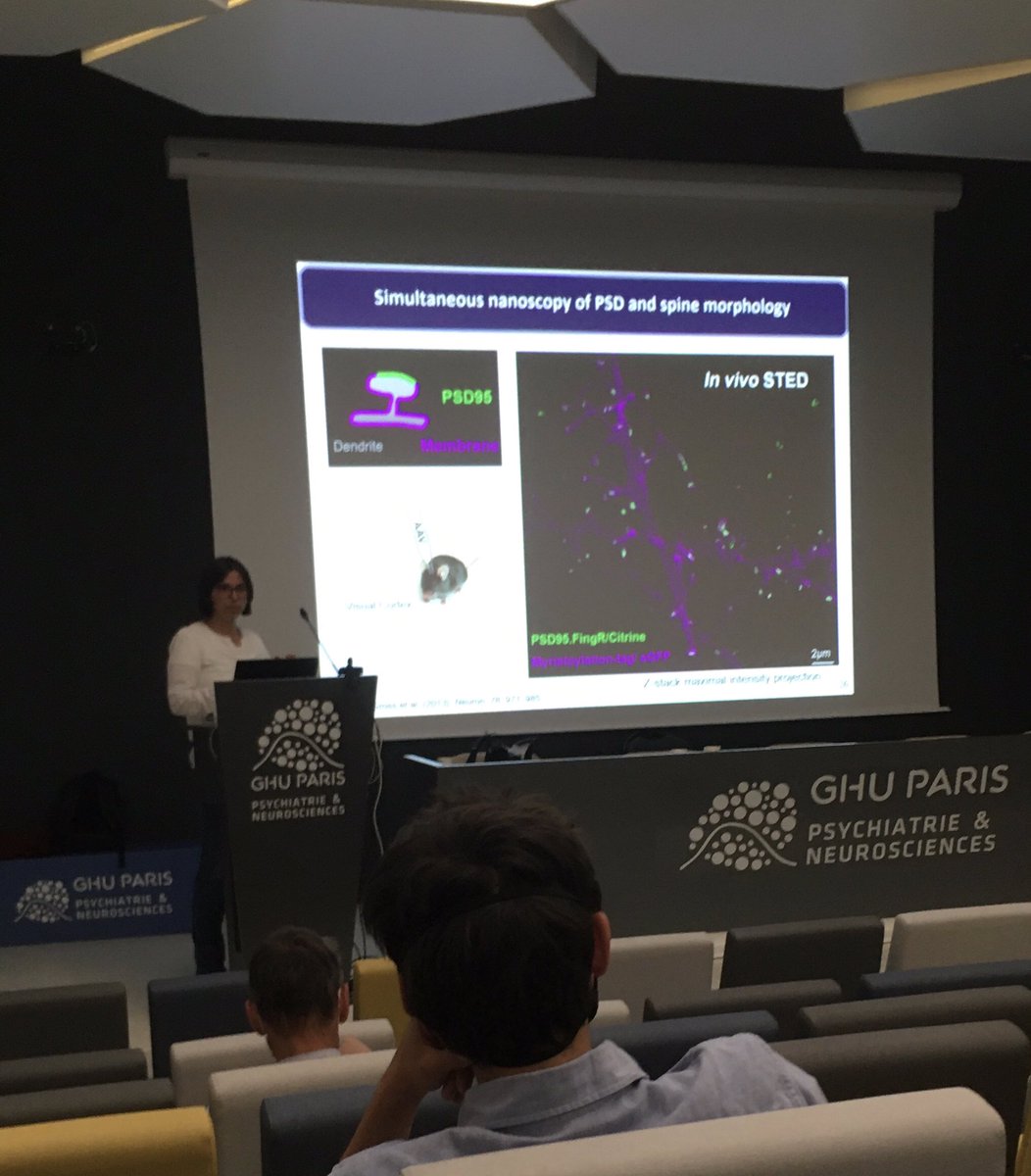
Very nice talk from @KatrinWillig about live sted in vivo microscopy of dendritic spine and PSD after enriched environnement learning at @GDR_ImaBio symposium @Insermu1266
Happy to share our recent work, pushing the limits of 3D resolution in fluorescence microscopy!
With special thanks to my colleagues and co-authors at @StefanHellLabs@JakobsLab@mpi_mr_hd and the IFNANO Göttingen.
@kirtiprakash25 @juliammichalska @PhotonPhilipp @HeintzmannLab@AgEggeling@LabSauer@LSchermelleh@Abberior To clearify, A is measured in vivo with Citrine, B in vitro with EGFP, live cell. This is about two-color STED of the living mouse brain. Showing also time lapse. Raw data, without deconvolution. doi: 10.7554/eLife.73603
Of course STED can go easily below 50 nm.
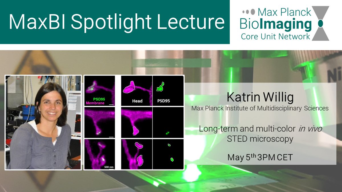
📢#Spotlightlecture with @KatrinWillig! She will present lots of fascinating data on in vivo #STED microscopy of #neurons. Examples of her recent works https://t.co/ES8UvfRtAY and https://t.co/dOjP7G3i5R
Join us on Zoom on May 5th ➡️https://t.co/DVK8Oo2R3M
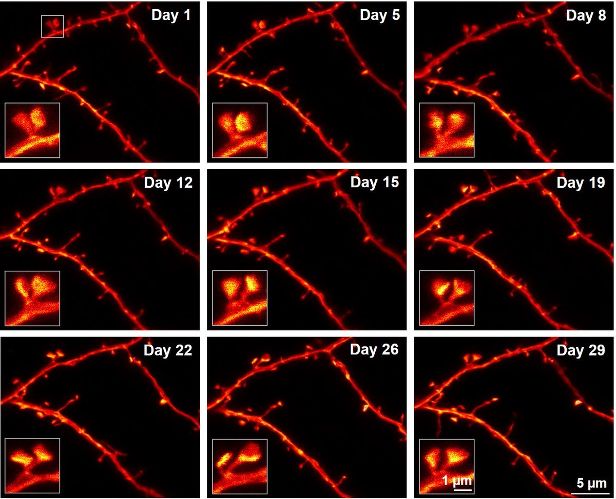
Happy to see that our paper is out in early view @eLife: We use two-color in vivo #superresolution#STED#microscopy to show the patterning and remodeling of the #synaptic nanoarchitecture after environmental enrichment.
https://t.co/H5HLRwpJJE
2. Activity-enhanced rearing in environmental enrichment decreases the variability of PSD95 assembly and spine head size; the size distributions are sharper than in controls raised in standard cages.
3. The nanostructure of PSD95 is more dynamic after environmental enrichment.