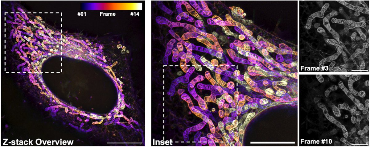
We got a belated Christmas present from @PNASNews: We made it onto the cover of the last issue for this year 😍 https://t.co/qjfToPXIlu
If you want to know more about the background, read our article about a new live-cell dye for mito imaging: https://t.co/mZ6BhnX7aE
Thrilled to announce that our work on a STED simulation software designed for the validation of AI approaches and training of RL agents for autonomous control of STED imaging parameters is out now @NatMachIntell!
@universitelaval@CERVO_ULaval@IID_ULaval
https://t.co/X9KK1DlPn4
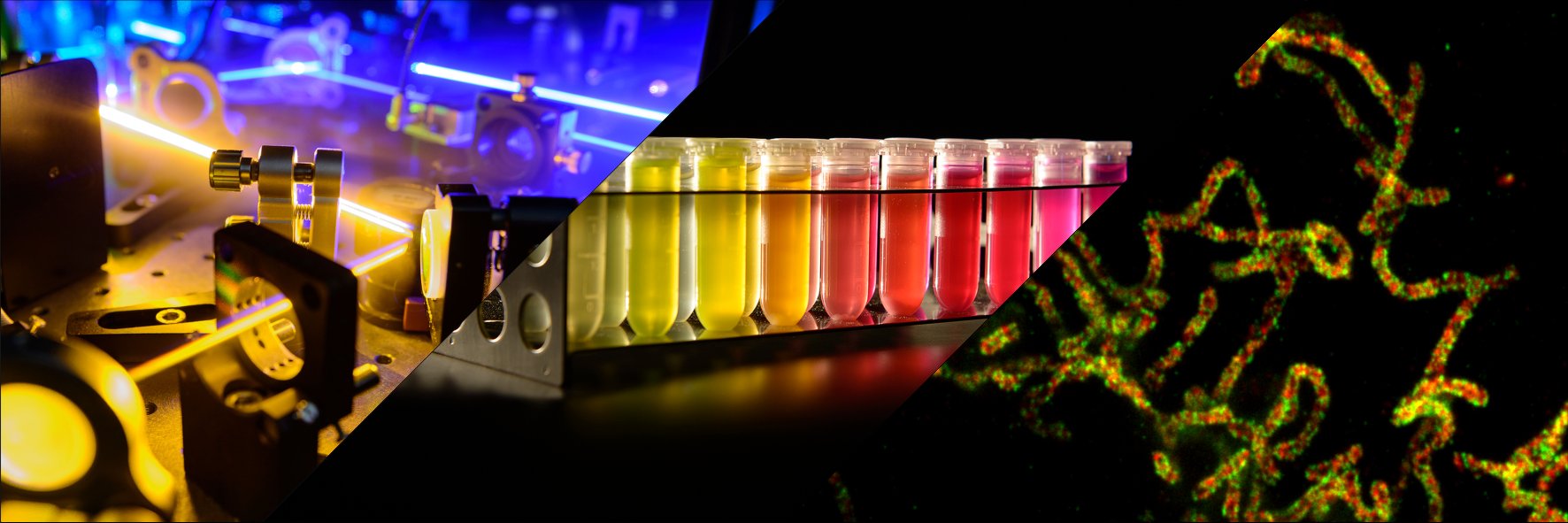
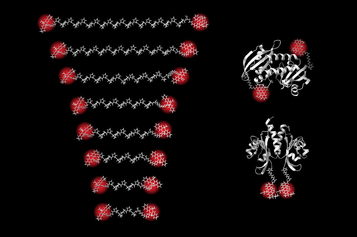
A team led by Steffen Sahl & @Stefan_W_Hell@mpi_nat and @mpi_mr_hd has succeeded in measuring distances within biomolecules using the #MINFLUX microscopy method, down to 1 nanometer and with Ångström precision. Read more in our press release:
https://t.co/NuAdZkXf6v
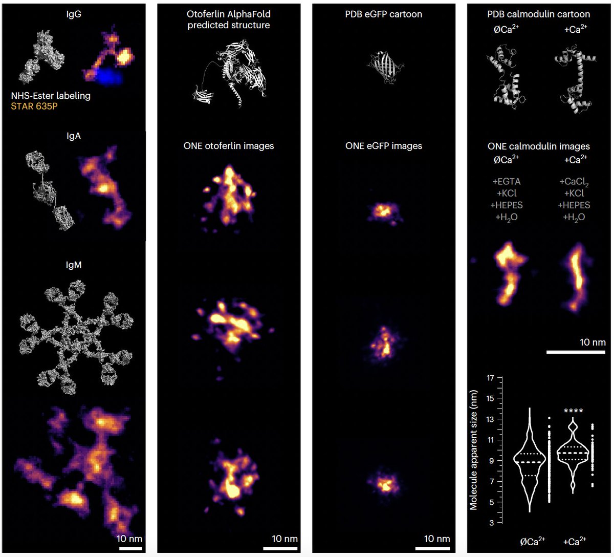
Protein shapes can now be studied using ONE🔬, a combination of ExM and #fluorescence fluctuation analysis. Happy to have contributed to this paper published in @NatureBiotech with many great scientists. Special thanks to @AliHShaib and Silvio Rizzoli. https://t.co/KKQw1ywZ2r
A nice talk from Stefan in PKU, with a lot of mitochondria, superresolution, and some twists to cryoEM in the end . Then we ended the day in the Emperior's diner for dinner😀 @JakobsLab@ZhixingChen2@xipeng1
Beautiful work on the in situ structure of ATP Synthase by @dietrich_th from the Kühlbrandt group published today in @ScienceMagazine 🔬 Discover how this important protein complex shapes the inner mitochondrial membrane! 🧪
https://t.co/xoRDR7zMgJ
Super excited to share our latest work on the native in-cell organization of the mitochondrial respiratory chain 🥳
Using cryo-electron tomography🔬, we show how the respiratory complexes (and other complexes) are organized inside native mitochondria!
https://t.co/cxHl1j20CJ
Want picture-perfect STED images of TOM & Co?
-> follow our detailed protocol out today in
Methods in Enzymology Vol. 710
"STED super-resolution microscopy of mitochondrial translocases"
Great work together with @InamdarKaushik , @cdanieljans. @JakobsLab
https://t.co/WOB7evRV4k
🚀Excited to share our latest paper, where we explore dynamic changes in mtDNA heteroplasmy in living cells! Discover how asymmetric partitioning and mitochondrial dynamics drive mtDNA variant segregation! 🔬https://t.co/2jDc93D4Sr #Mitochondria#microscopy#microfluidics#mtDNA
GBM Compact: Focus on Imaging Frankfurt, September 26-27, 2024 Deadline for early registration and abstract submission: June 30
https://t.co/VKpLp7KHkm
👇 👇 👇 👇 👇
The GBM Compact conference „Focus on Imaging“ takes place from September 26-27 in Frankfurt! Early registration+abstract submisison is possible until June 30. Please visit the conference website for registration and further information: https://t.co/VKpLp7KHkm
Are you interested in the intricate biology of fluorescent proteins? Do you want to push the power of MINFLUX microscopy even further? Live-cell imaging of mitochondrial protein dynamics tickles your brain?
We want you 🧐
Check out the details below 👇
https://t.co/yBQGEutzyI
Here comes our next gen PK Mito Orange Fix that can be fixed to IM for you to scrutinize the wonderland of mitochondria under STED, in a multiplexed way!
Many thanks for the growing PK Mito team! @stephan_till@JakobsLab and Christian Juengst!
https://t.co/53mujZAhZm
After countless requests, we're thrilled to introduce PK Mito Orange FX - a fixable variant of PKMO. PKMO FX allows immunolabeling and CLEM approaches.
Happy to have contributed to this project led by @ZhixingChen2 and Christian Jüngst.
#mitochondria
https://t.co/nC6gX3LBGt
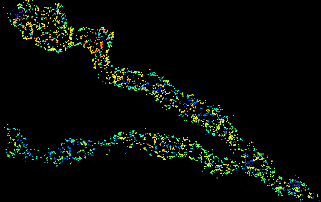
WHAT % OF YOUR TARGET BIOMOLECULES ARE YOU REALLY IMAGING?
The *labeling efficiency* of your experiment is extremely important for image quality and quantitative results in super-resolution!
Super excited about our new article in @naturemethods! 🤩
https://t.co/QuVsvRFnRE