“Edematoso pero intravascularmente seco” sí tiene una explicación fisiológica.
En sepsis: hipovolemia + hipoalbuminemia + edema → no es solo “fuga capilar”, también hay ↓ retorno linfático.
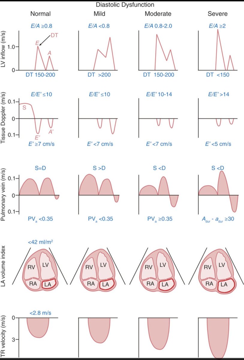
Understanding Diastolic Dysfunction with Doppler Echocardiography
This chart beautifully compares Doppler findings in normal vs. varying grades of LV diastolic dysfunction from inflow (E/A, DT) to tissue Doppler (E′/A′), pulmonary vein flow, LA volume, and TR velocity.
Source: Catherine M. Otto, Textbook of Clinical Echocardiography
#POCUS#echofirst#Nephpearls
RVOT pulse-wave Doppler can provide useful clues about pulmonary vascular resistance.
In normal individuals (A), the waveform has a smooth, dome-shaped appearance, with peak velocity occurring in mid-systole, reflecting a compliant, low-resistance pulmonary circulation.
As RV afterload increases, the waveform gradually becomes more triangular. The RVOT acceleration time shortens, and the peak velocity shifts earlier into systole (B).
With further increases in pulmonary vascular impedance and reduced arterial compliance, a characteristic mid-systolic notch may appear (C), creating the classic "W sign."
In advanced pulmonary hypertension with RV failure, the Doppler envelope becomes smaller and more abbreviated, with a very short and steep AccT (D). This reflects rapid equilibration of RVOT and proximal pulmonary artery pressures due to severe afterload elevation.
Like most POCUS findings, RVOT Doppler should be interpreted in the context of the overall echocardiographic picture rather than in isolation.