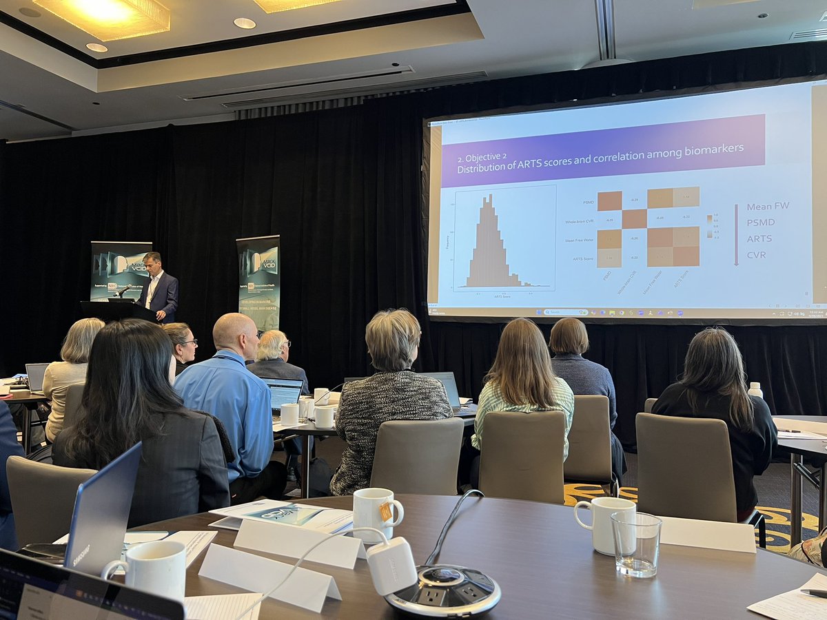
Dr. Arnold Evia presenting MARBLE, our novel in-vivo marker of LATE-NC #AAIC25 This is the result of machine learning, ex-vivo MRI, in-vivo MRI, pathology data, and a lot of hard work from our amazing interdisciplinary team @alzassociation@rushalzheimers@IITEngineering
The most comprehensive and conclusive paper on how different neuropathologies impact the volume and shape of different deep gray matter structures in older adults is now published on Human Brain Mapping https://t.co/vDjTnO5wXB @IITEngineering@rushalzheimers
🚨 Excited to share my first-author paper from @MRIatIIT, now published in Human Brain Mapping 🧠
We analyzed 842 autopsied older adultsusing ex vivo MRI + neuropathology to uncover how age-related pathologies independently impact the volume & shape of deep gray matter structures.
📍Regions: Hippocampus, Amygdala, Thalamus, Caudate, Accumbens, Putamen
🧬Pathologies: #AD #Tangles #AmyloidBeta #TDP43 #LATE #LewyBodies #Atherosclerosis #Arteriolosclerosis #Infarcts #Microinfarcts #CAA
🙏Grateful to my co-authors & collaborators at @rushalzheimers@IITEngineering, esp. Dr. Konstantinos Arfanakis
📖Read: https://t.co/SK69BMg8lI
#Neuroimaging #Alzheimers #MRI #BrainMapping #Neuropathology #SubcorticalStructures
Exciting updates on the ARTS biomarker today at #MarkVCID!
Developed at @MRIIT, ARTS is an MRI-based, fully automated tool detecting brain arteriolosclerosis. Dr. Arfanakis presented new findings: higher ARTS scores are associated with greater vascular risk, worse executive function, and cognitive decline. Stronger associations observed in females and Black participants.
Promising steps toward non-invasive vascular brain health biomarkers! #MRI #Neuroimaging #Biomarkers #BrainHealth
Excited to be at the 2025 #MarkVCID Annual Conference in Chicago! Looking forward to updates on imaging biomarkers (MRI ARTS, CVR, Free Water, PSMD) and plasma NfL. Great to learn from leaders advancing vascular cognitive impairment research! 🧠✨ #BrainHealth#DementiaResearch
We now have a blood biomarker that is even more strongly associated with tau PET and cognitive symptoms than even p-tau217! eMTBR-tau243 will help us to understand whether cognitive impairment is likely to be related to Alzheimer's pathology. #ENDALZ
https://t.co/TX36rUjgjp
Hippocampal neuronal loss (HNL) plays a critical role in cognitive decline—especially in LATE-NC, while ADNC-related decline follows a different trajectory. 🧠📉 This study also reinforces what we observe in our lab at @MRIatIIT : LATE-NC has a distinct neurodegenerative signature, with HNL acting as a major mediator of cognitive impairment. These insights reshape our understanding of brain aging and could drive more targeted interventions for Alzheimer's and LATE. The future of neurodegeneration research lies in decoding these mechanistic differences! 🔬✨ #Neuroimaging #Alzheimers #LATE #CognitiveDecline #BrainHealth
Check out our new paper with @JASchneiderMD@beyoung40 and @rushalzheimers colleagues.
Hippocampal neuronal loss is a prominent feature of LATE-NC and contributes to cognitive decline. - Agrawal - Alzheimer's & Dementia - Wiley Online Library https://t.co/iD9EQsDrDC
From my journey—starting in Electrical Engineering and transitioning into Biomedical AI and Neuroimaging—I've learned some invaluable lessons:
1️⃣ Interdisciplinary Knowledge is Powerful 🚀
2️⃣ Hands-on Work Beats Theory Alone 🔬
3️⃣ Patience and Perseverance Pay Off 🧠
4️⃣ Networking & Sharing Work Matters 🌎
5️⃣ AI in Healthcare is the Future 🔥
💡 If you’re on a similar path—whether in AI, neuroscience, or medical imaging—keep learning, experimenting, and sharing your work! 🚀
#AI #MachineLearning #Neuroimaging #Alzheimers #BiomedicalEngineering #PhD #Research
Attention has been the key component for most advances in LLMs, but it can’t scale to long context. Does this mean we need to find an alternative?
Presenting Titans: a new architecture with attention and a meta in-context memory that learns how to memorize at test time. Titans are more effective than Transformers and modern linear RNNs, and can effectively scale to larger than 2M context window, with better performance than ultra-large models (e.g., GPT4, Llama3-80B).
For anyone who doesn't know about it (and if you have a long commute like me!), I strongly recommend Peter Bandettini's Neurosalience podcast - he interviews many key people in neuroimaging and neuroscience on their careers, interests, key findings, what motivates them. I once binge-listened to it while driving for 10 hours from Glasgow to London via Yorkshire :)
PhD Students - Identifying research gap is one of the biggest challenges.
How to identify the research gap for your PhD?
Here is a stepwise process that can help you to identify gap for your PhD.
𝟏. 𝐈𝐝𝐞𝐧𝐭𝐢𝐟𝐲 𝐫𝐞𝐬𝐞𝐚𝐫𝐜𝐡 𝐚𝐫𝐞𝐚:
Before identifying the gap, you need to identify the area. This is quite easy. The area can either come from your previous interests or your supervisors can give it to you. For example, detecting cyber-attacks is a research area.
𝟐. 𝐑𝐞𝐚𝐝 𝟓-𝟏𝟎 𝐥𝐢𝐭𝐞𝐫𝐚𝐭𝐮𝐫𝐞 𝐫𝐞𝐯𝐢𝐞𝐰𝐬 𝐢𝐧 𝐭𝐡𝐞 𝐚𝐫𝐞𝐚:
Once the area is identified, search for 5-10 most relevant literature reviews/secondary studies in the area. These papers have already reported a summarized view of the existing primary studies. Read these papers carefully to understand what literature already exists in the area.
𝟑. 𝐅𝐨𝐜𝐮𝐬 𝐨𝐧 𝐭𝐡𝐞 𝐟𝐮𝐭𝐮𝐫𝐞 𝐫𝐞𝐬𝐞𝐚𝐫𝐜𝐡 𝐢𝐧 𝐭𝐡𝐞 𝐥𝐢𝐭𝐞𝐫𝐚𝐭𝐮𝐫𝐞 𝐫𝐞𝐯𝐢𝐞𝐰𝐬:
While reading these 5-10 literature reviews, focus on the future research areas, open challenges, and discussion section. Identify 3-5 research directions from these literature reviews. Detecting data exfiltration attacks is a research direction.
𝟒. 𝐂𝐡𝐞𝐜𝐤 𝐞𝐱𝐢𝐬𝐭𝐢𝐧𝐠 𝐥𝐢𝐭𝐞𝐫𝐚𝐭𝐮𝐫𝐞 𝐫𝐞𝐥𝐚𝐭𝐞𝐝 𝐭𝐨 𝐭𝐡𝐞 𝐢𝐝𝐞𝐧𝐭𝐢𝐟𝐢𝐞𝐝 𝐝𝐢𝐫𝐞𝐜𝐭𝐢𝐨𝐧𝐬:
Just to make sure that you don't end up doing something that already exists, search primary studies related to the research directions. Drop the ones where exactly similar works exist.
𝟓. 𝐃𝐢𝐬𝐜𝐮𝐬𝐬 𝐭𝐡𝐞 𝐢𝐝𝐞𝐧𝐭𝐢𝐟𝐢𝐞𝐝 𝐝𝐢𝐫𝐞𝐜𝐭𝐢𝐨𝐧𝐬 𝐰𝐢𝐭𝐡 𝐲𝐨𝐮𝐫 𝐬𝐮𝐩𝐞𝐫𝐯𝐢𝐬𝐨𝐫𝐬:
Make a few slides to present the remaining directions to your supervisors. From here, you should pick the direction where you and your supervisor see the most potential.
𝟔. 𝐂𝐨𝐧𝐝𝐮𝐜𝐭 𝐚 𝐥𝐢𝐭𝐞𝐫𝐚𝐭𝐮𝐫𝐞 𝐫𝐞𝐯𝐢𝐞𝐰 𝐨𝐧 𝐭𝐡𝐞 𝐢𝐝𝐞𝐧𝐭𝐢𝐟𝐢𝐞𝐝 𝐝𝐢𝐫𝐞𝐜𝐭𝐢𝐨𝐧:
Once the direction is picked, do a literature review on the specific direction. If no paper exists at all in this direction, this could mean two things - either the topic is not worth doing research or the topic is good but too new.
𝟕. 𝐈𝐝𝐞𝐧𝐭𝐢𝐟𝐲 𝐜𝐫𝐢𝐩𝐬 𝐠𝐚𝐩𝐬 𝐯𝐢𝐚 𝐭𝐡𝐞 𝐥𝐢𝐭𝐞𝐫𝐚𝐭𝐮𝐫𝐞 𝐫𝐞𝐯𝐢𝐞𝐰:
This literature review process should get you the crisp gap. However, it won't come automatically. While reading each paper, note down the points that you think could be worth future research. This will become part of your discussion or future research section. For example, detecting data exfiltration attacks in real-time is a gap.
𝟖. 𝐂𝐡𝐞𝐜𝐤 𝐫𝐞𝐬𝐨𝐮𝐫𝐜𝐞𝐬 𝐫𝐞𝐪𝐮𝐢𝐫𝐞𝐝 𝐭𝐨 𝐟𝐢𝐥𝐥 𝐭𝐡𝐞 𝐠𝐚𝐩:
Once you have identified the research gap, check what kind of resources, data, infrastructure, etc, you need to conduct this research. Make sure that you can have access to these resources before you start working on the gap. Anything to be added?
You can now join our new LATE @ISTAART PIA! 🧠
https://t.co/lQ3Ny5VJV8
With this PIA, we want to raise awareness of this dementia-driving pathology, foster collaborations, and ultimately improve TDP-43 targeting therapies.
Stay tuned!
#dementia
Excited to present “Brain Atrophy in Alzheimer’s and LATE Neuropathology” (#T2-SPNR-11) at #RSNA24!
📅 Dec 3, 2024
🕘 9:00-9:30 AM
📍 Learning Center, Hall D
Join me to discuss Alzheimer’s and neuroimaging! #AlzheimersResearch#LATE#RSNA#RSNA2024
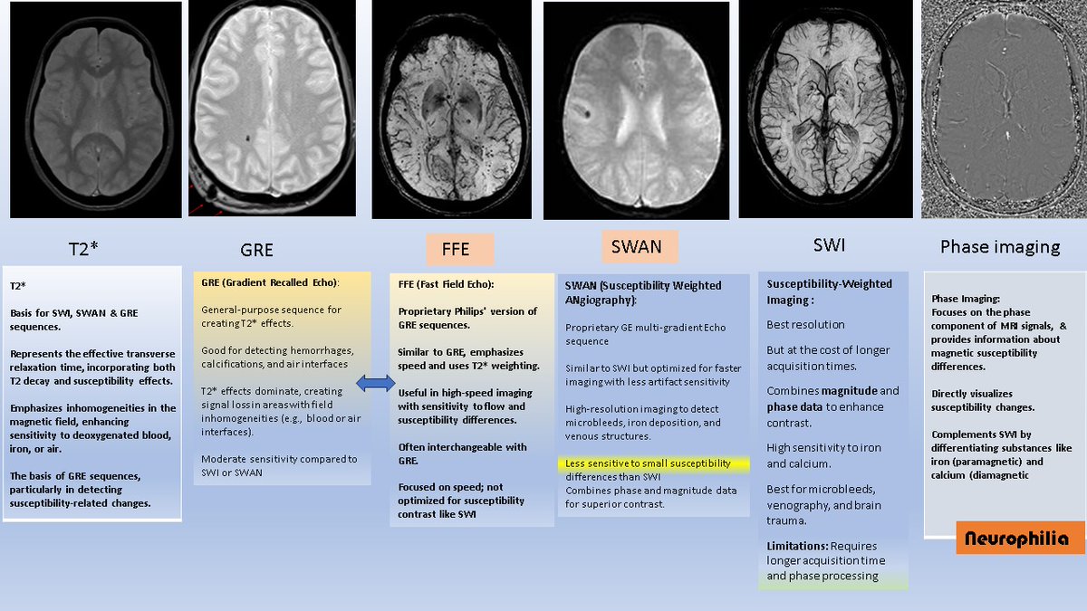
MRI for the non-radiologists. Do the terms FFE, GRE, T2*, SWI, SWAN & Phase imaging confuse you? Here is a handy reference sheet for that. Without going into any of the physics of MRI, the 1st point to understand is;
1. T2* (pronounced "T2-star") is the basis of other sequences, such as SWAN, GRE, and FFE
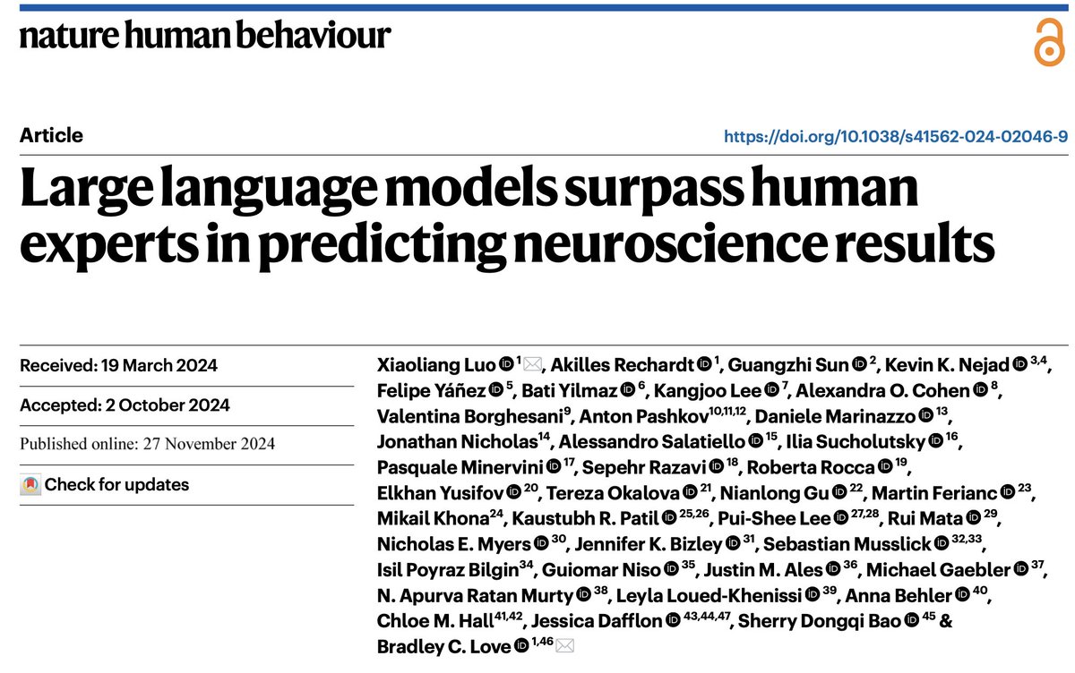
"Large language models surpass human experts in predicting neuroscience results" w @ken_lxl and https://t.co/YOhCmQlJsu. LLMs integrate a noisy yet interrelated scientific literature to forecast outcomes. https://t.co/49WYirBdBv 1/8
Great to see our #MIITRA atlas tested at FMRIB. The image quality of #MIITRA tops the list of atlases of the older adult brain! This is in agreement with our findings. The newer version of #MIITRA is even better https://t.co/QMDuPzmyPA @NIHAging@NACCData https://t.co/TOKmOZBd40