New in AJO: Can we risk-stratify high-risk PDR with a single widefield OCTA scan? Our work at @CaseyEye, an amazing engineer–retina clinician collaboration, shows elevated RNV at baseline was strongly associated with VH/TRD.
https://t.co/Cj0T8fIGah
Yali Jia @YaliJiaOCT et al. from Casey Eye Institute @CaseyEye quantify choriocapillaris flow deficits using projection-resolved optical coherence tomographic angiography and evaluate whether they are correlated with diabetic retinopathy.
https://t.co/O0oycdtSHj
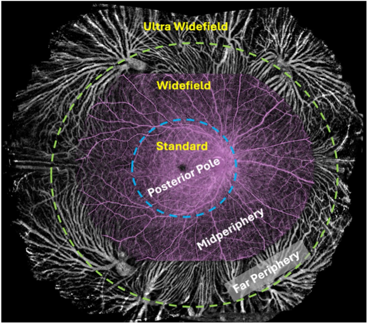
@CaseyEye’s fresh review paper “Widefield OCT Angiography” available on PRER Journal. Widefield guideline, technical innovations, clinical utility and more….https://t.co/IvBBiNgRum
@CaseyEye’s fresh review paper “Widefield OCT Angiography” available on PRER Journal. Widefield guideline, technical innovations, clinical utility and more….https://t.co/IvBBiNgRum
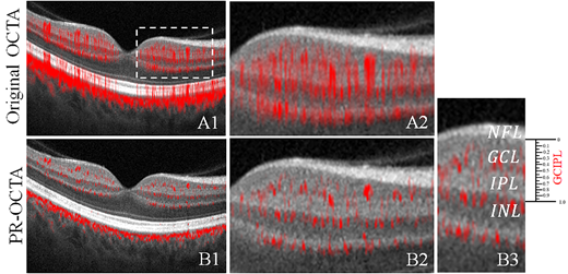
In the New Frontiers in Optical Coherence Tomography Issue https://t.co/x8eJaF06aP, David Huang et al. @CaseyEye study the Anatomy of the Macular Ganglion Cell Layer Plexus on Projection-Resolved Optical Coherence Tomographic Angiography, https://t.co/yj6cBp63C6.
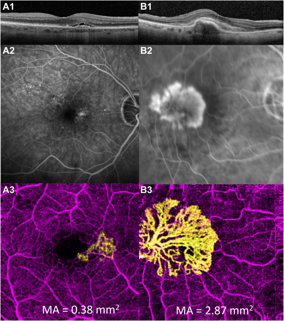
AI algorithm combines 3D structural OCT and en face OCTA simultaneously to improve accuracy of macular neovascularization segmentation and detection.
#NEIfunded@CaseyEye@MahidolU@SirirajMedicine @YaliJiaOCT
@AAOJournal: https://t.co/Qt43cekdnb
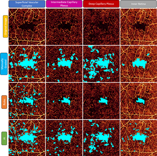
Yali Jia et al. present their work on Nonperfused Retinal Capillaries—A New Method Developed on OCT and OCTA, https://t.co/AuJdPgS3QU.
@YaliJiaOCT @CaseyEye@OHSUNews
Researchers find AI algorithm for quantifying retinal nonperfusion area based on OCTA in diabetic retinopathy is a promising tool for automated screening and diagnosis. @ARVOiovs@YaliJiaOCT https://t.co/uShxS2sXgM