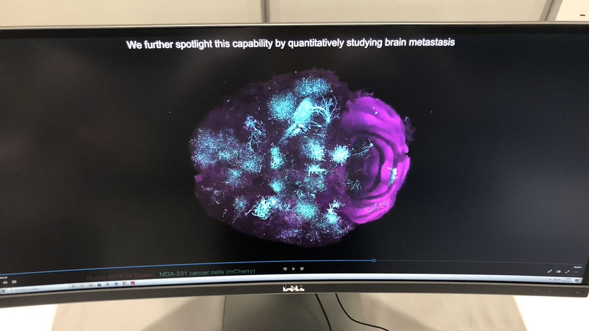
Calling all pathology enthusiasts! We're presenting at USCAP, will you be there? Don't miss @RederredeR present about next generation cancer diagnostics and the role 3D pathology will play. We can't wait to share what we have been up to with AI-powered #3Dspatialbiology
We are proud to be featured in @wrfseattle 2021 year in review, the support from WRF and WRF Capital has been invaluable. Here is to 40 more years with our incredible partners!
We're #hiring! Help us transform drug development with #3Dspatialbiology and lead an incredible team of researchers! Apply here: https://t.co/nGOHyd8O5V
An important new paper by @jonliu123 and team demonstrates the advantages of 3D imaging and deep learning over traditional 2D biopsy slide exams when it comes to diagnosing prostate cancer. Press release from @uwnews: https://t.co/R83Px6BoCm @uwmnewsroom@cwru@lightspeedmicro
An important new paper by @jonliu123 and team demonstrates the advantages of 3D imaging and deep learning over traditional 2D biopsy slide exams when it comes to diagnosing prostate cancer. Press release from @uwnews: https://t.co/R83Px6BoCm @uwmnewsroom@cwru@lightspeedmicro
Excited to share an updated preprint on our new hybrid open-top light-sheet microscope. It has been a blast collecting data over the last year with our most powerful and versatile system to date. Big thank you to @jonliu123 and all of our co-authors! https://t.co/2935CuoQs3
Proud of Weisi Xie for leading this study/preprint. Special thanks to L. True, @anantm, @RederredeR, @adam_k_glaser, & C. Koyuncu. Prostate cancer risk stratification via non-destructive 3D pathology with annotation-free gland segmentation and analysis https://t.co/UE2RaaZjIi
I am excited to announce our $4M financing round, led by Dynamk Capital! We look forward to growing our team, enhancing our technology, and continuing to provide biological insights for our Pharma customers using our end-to-end 3D pathology platform.
https://t.co/TZfwG15OdQ
First human brain slice imaging results. 2 micron isotropic raw data, but fused with 8x down-sampling to render this video. Autofluorescence background with amyloid plaques in green!
Excited to share our vision for nondestructive 3D pathology for clinical decision support! We discuss optical, computational and translational challenges. https://t.co/qVCHAXjQ39 @adam_k_glaser@hopelessk@RederredeR@UWMadisonLOCI@anantm
Check out this video from @adam_k_glaser showing how open-top #lightsheet microscopes will change the way we process and view tissues. Watch the microcope in action at 0:23 and dive through the tissue samples at 0:35.
Excited to share our new multi-immersion open-top light-sheet microscope https://t.co/HCnrAvj2jl. Simple mounting and versatile imaging of multiple specimens cleared with a variety of protocols. Thanks to all of our collaborators. @jonliu123@RederredeR @Ti_Sapph @jsdaniel02