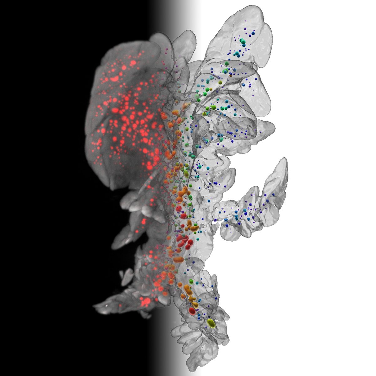
After a difficult time, we’re back on track and our 3rd manuscript of the year has been accepted for publication! 😀 A great collaboration with @lighttodiabetes & @pthorn14 “Insulin-binding peptide probes provide a novel strategy for pancreatic β-cell imaging” @ACSBioMed
Our new research paper is out in eNeuro: Distinct Opsin 3 (Opn3) Expression in the Developing Nervous System during Mammalian Embryogenesis https://t.co/huGE2DC0DX
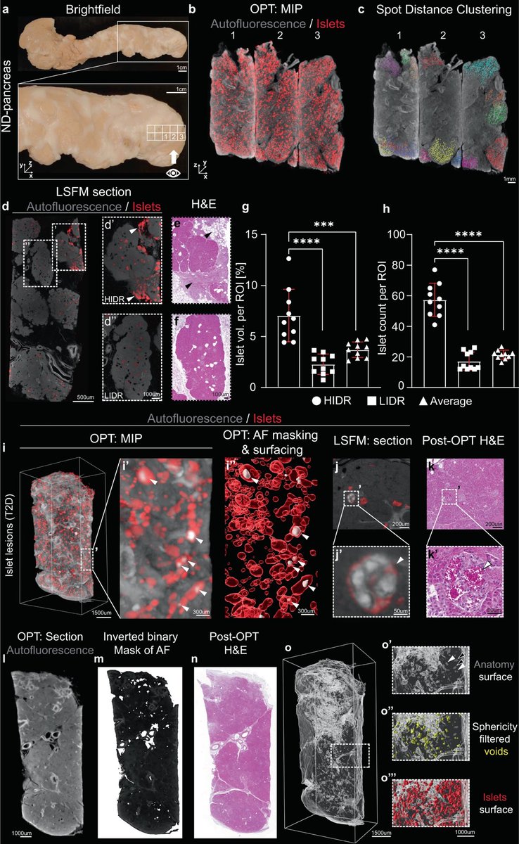
In this study, Hahn et al. present a method for visualizing the endocrine human pancreas in 3D and apply it to individuals with type 2 diabetes to reveal morphological differences in the endocrine pancreas. @lighttodiabetes@UmeaFor https://t.co/jSnmohM1wq
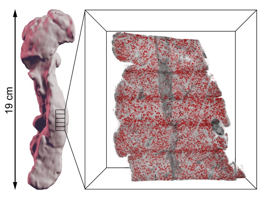
Our article is out @CommsBio! By the use of a 3D printed matrix we show how human organs (in our case the pancreas) can be specifically labelled, imaged by optical mesoscopic imaging techniques & reconstructed back together in 3D. https://t.co/1CP4RZZGqz
#LSFM#imaging@UmeaFor
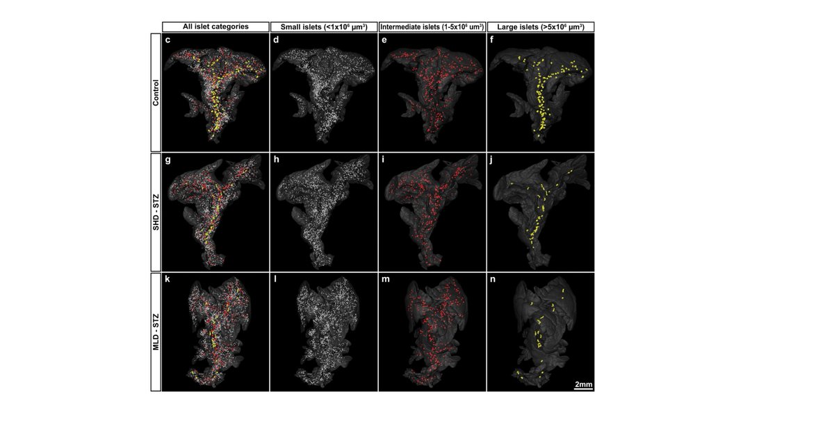
Streptozotocin-induced hyperglycemia primarily results from beta-cell dysfunction rather than ablatation in mice. This study provides insight into how the most widely used preclinical diabetes model works https://t.co/npOLRNcaZk
Another paper out! Rapid Autofluorescence based 3D imaging of #PancreaticCancer . The video shows a human pancreatic cyst with vessels (red) and islets of Langerhans (green) imaged with the Ultramicroscope II.
https://t.co/KuJyqCPezY
Congratulations Professor Emmanuelle Charpentier for the award of the 2020 Nobel Prize in Chemistry for the discovery of the gene-editing tool CRISPR-Cas9.
#EmmanuelleCharpentier#NobelPrize
https://t.co/ijILdp1RUm
🔎 Researchers from @UmeaUniversity show that hyperglycemic #Streptozotocin-treated mice still harbor a large pool of remaining β-cells but display pancreas-wide downregulation of glucose transporter type 2.
🔓 Open access https://t.co/umKiVL6wV4
The video depicts a diabetic mouse pancreas and its different lobes (splenic lobe in green, gastric lobe in blue and duodenal lobe in yellow) imaged by Optical Projection Tomography. The surviving islets of Langerhans (in red) are dysfunctional.
https://t.co/xysdWRREod
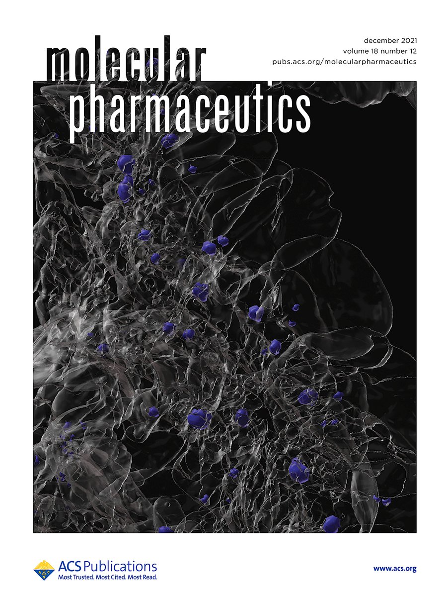
Breaking news! UCMM would like to congratulate Prof. Ulf Ahlgren’s group on their excellent new paper published yesterday in Communications Biology (https://t.co/rdNWfGKXtZ). The image depicts a mouse pancreas imaged by Optical Projection Tomography (OPT).
Now published! A new study in Nature's @CommsBio on the Streptozotocin diabetes mouse model, the most used diabetes model in rodents. Using advanced imaging, we show that the majority of islets remain, but surviving islets of Langerhans are dysfunctional.
https://t.co/xysdWRREod
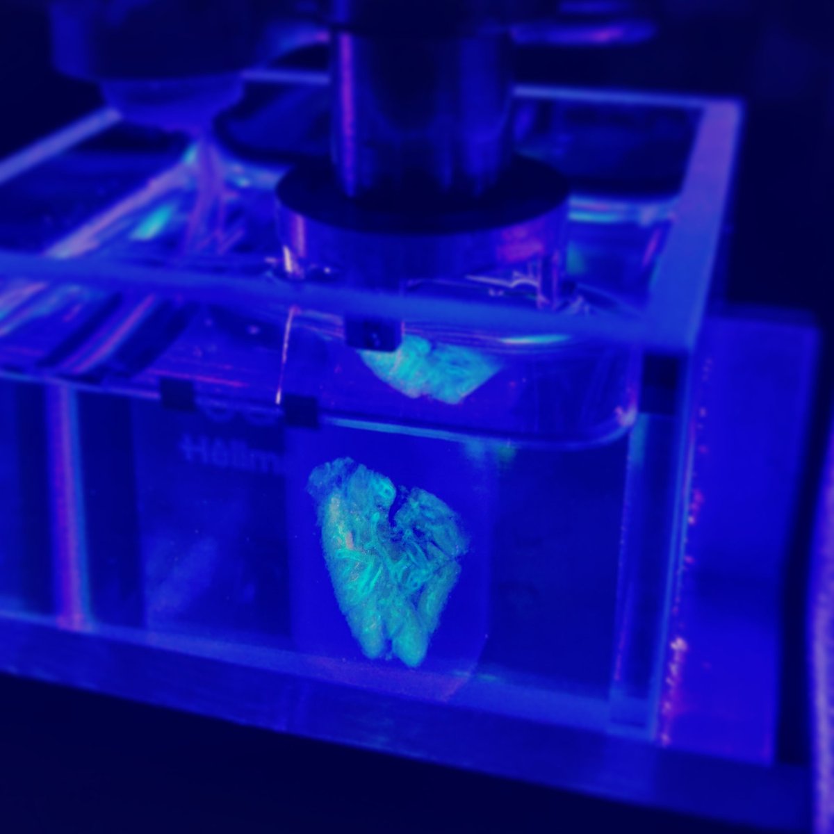
🔬Check out this stunning fluorescent slab of human pancreas in our 3D Microscope (Optical Projection Tomography). We make the sample transparent by adapting the refractive index, which allows to shine through the sample.
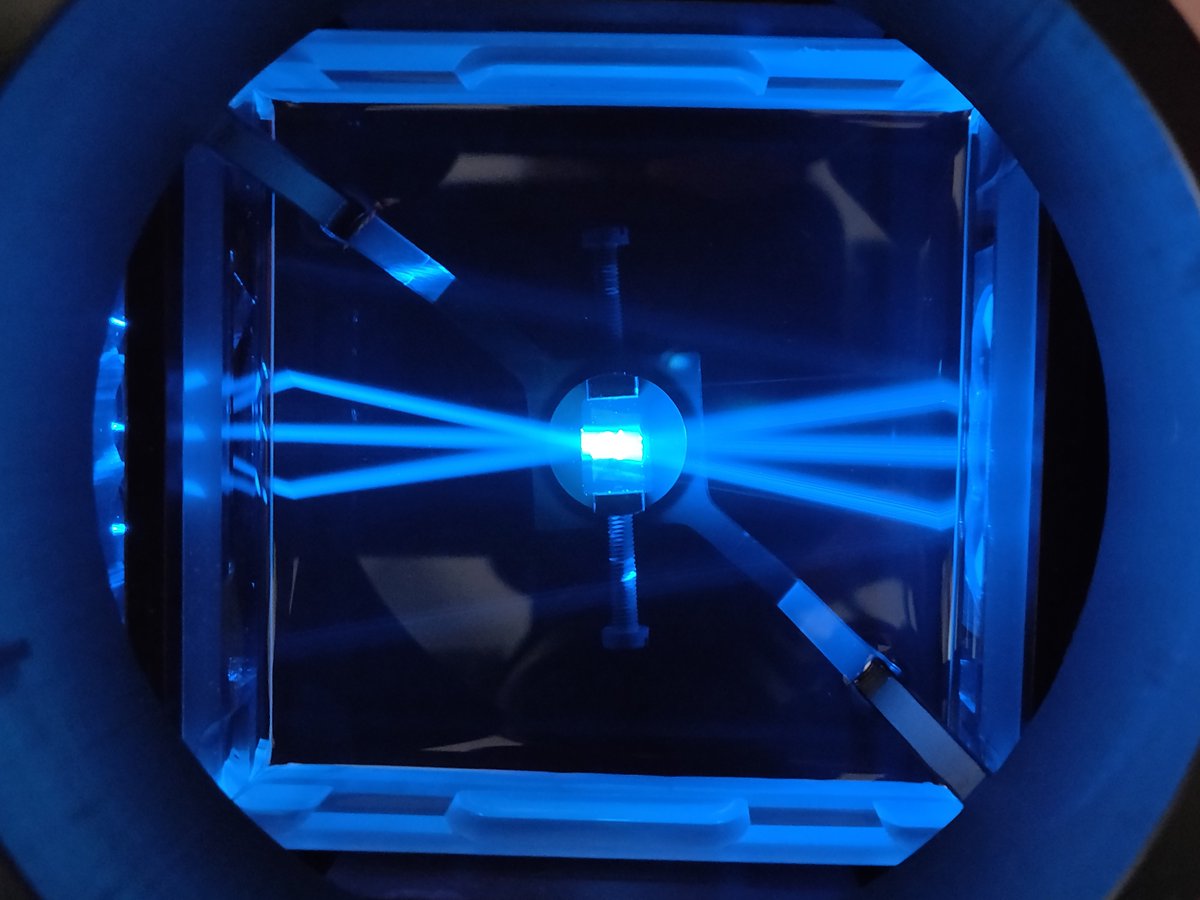
Bringing light to diabetes on world diabetes day: 6 Lasers are used to analyse a pancreatic specimen in a Lavision Biotec 2nd generation Ultramicroscope.