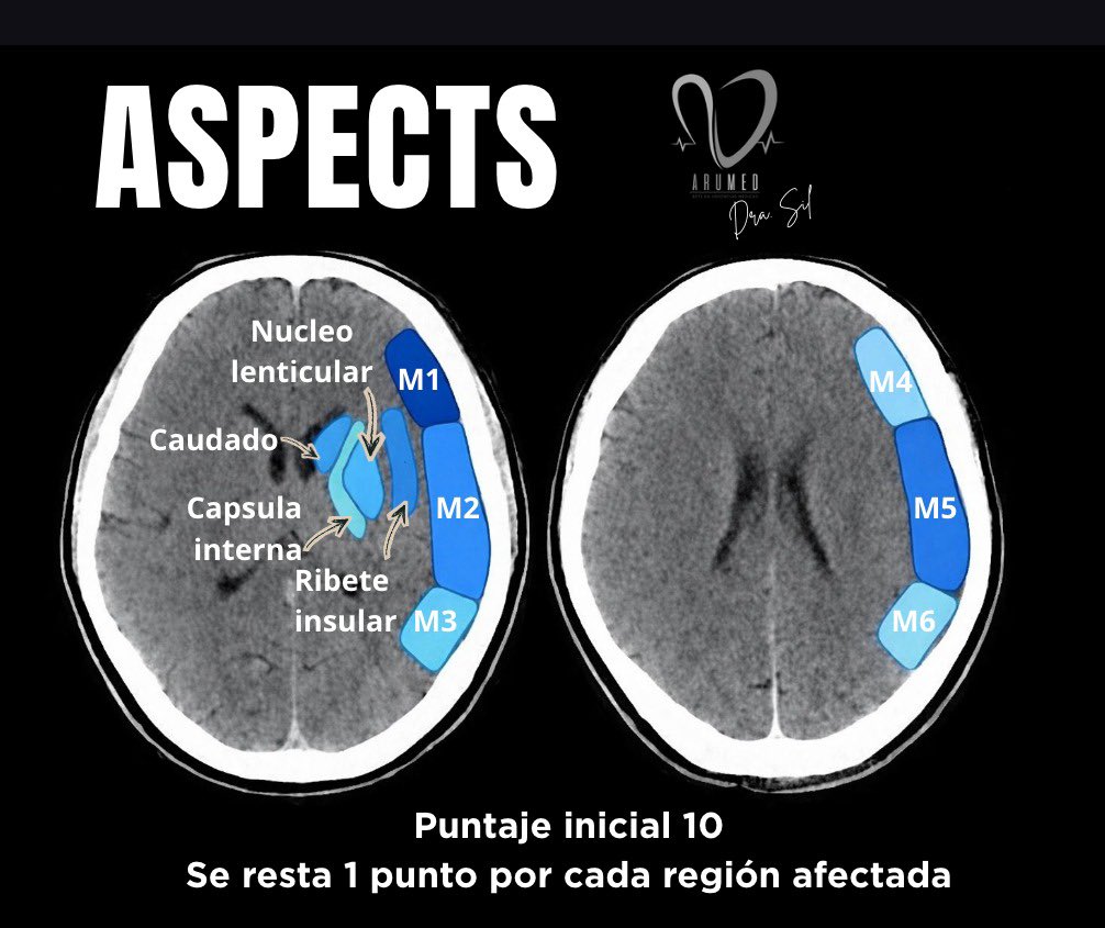
ASPECTS: decide rápido, trata mejor
Alberta Stroke Program Early CT Score evalúa cambios isquémicos tempranos en TAC simple en territorio de la arteria cerebral media (ACM) en EVC
Puntaje inicial: 10
➖ 1 punto por cada región afectada
An easy way to differentiate these midbrain syndromes is to remember that, moving from anterior to posterior, all three involve oculomotor nerve damage, with additional distinct features:
Weber’s syndrome – affects the anterior midbrain, involving the corticospinal tracts, leading to contralateral weakness.
Benedikt’s syndrome – involves the mid-midbrain, including the red nucleus, causing abnormal involuntary movements such as tremor and chorea.
Claude’s syndrome – localized to the posterior midbrain, where superior cerebellar peduncle fibers (after decussation) are affected, resulting in cerebellar signs (think Claude = Cerebellar).
#Neurotip
Need help reading spine imaging? I’ve got your back!
It’s as easy as ABC!
This post is about an easy mnemonic you can use on every single spine study you see to increase your speed & make sure you never miss a thing!
Just remember ABCD:
A = Alignment
(1) look for unstable injuries
(2) look for malalignment that causes early degenerative change
B = Bones
On CT, look for fractures
On MRI, look for marrow lesions/edema
C = Cord/Canal
On CT, look at the canal contents for large masses or collections
On MRI, look for canal narrowing & cord edema
D= Discs/Degenerative
--Normal discs should look like a kidney on it’s side on axial images, w/tiny hilum/. Loss of this hilum means there a bulge
--On sagittal images, normal discs should look like jelly donuts. If they look like pancakes instead of jelly donuts, they are degenerated
Disc nomenclature:
Bulge = gaining weight & loosening belt (annulus gets loose)
Protrusion = hernia (annulus tears & disc protrudes through)
Extrusion = disc becomes like toothpaste & squeezes around everywhere
Free Fragment = like toothpaste on the toothbrush—completely separate from the disc
Now you know how to approach spine imaging studies in a systematic way! Hopefully, now reading spine imaging won’t be such back breaking work!
1/Feel like a fish out of water when it comes to water on the brain?
Read on for this month’s @Radiographics summary of what you need to know about hydrocephalus!!
https://t.co/wAJWQDOPWr
@cookyscan1@RadG_editor#RGphx
Say bye to questions about bilateral thalamic lesions!
Feeling bipolar about bilateral thalamic lesions?
Unfortunately, the differential for bilateral thalamic lesions isn’t binary!
But here’s an easy mnemonic that can help you to remember: THALAMIC!
Here's how it works:
T = Tumor (glioma)
H = Hypoxic/ischemic encephalopathy
A = Artery of Percheron Infarct
L = Loss of thiamine (Wernicke’s)
A = ADEM
M = Metabolic/Toxic
I = Internal cerebral vein thrombosis. Infection (west nile)
C = Creutzfeld-Jacob
Now you’ll never bypass a bilateral thalamic diagnosis with this easy mnemonic!