Microscopy core facility at the Douglas Hospital Research Centre (McGill University). Supports neuroscientists in their imaging needs. Photo Credit: @labriedion
Microscopy Tweeps! I have condensation on the chip of one of my Hamamatsu cameras...
Besides better regulating room temp and humidity, anyone has any suggestions on how to deal with this problem ?
🤔
Thanks for everyone’s feedback! So follow-up: Hamamatsu confirms it’s a seal problem from that particular batch of cameras. We are shipping it to have it repaired or replaced. 👍🏼
The STM Working Group on Image Alteration and Duplication Detection have developed best practice recommendations for handling image integrity issues
At our upcoming free to attend webinar, these draft recommendations will be presented
Register via: https://t.co/JAIxlDGafr
To all our users using @QuPath, learn the importance of acknowledging the hard often unrecognized work that goes behind writing such a tool AND in this case the struggle to release it open-source so we could all enjoy its use. Thank you @petebankhead and all your team!
A small number of people know the real background story to @QuPath, but most don't.
I didn't plan to ever tell it publicly, until a Google Alert today caught my eye.
A thread about open science & academia 👇 (1/n)
Let's celebrate #FluorescenceFriday with a look at intracranial lymphatic vessels in #zebrafish Qdot angiography in magenta, lymphatics and FGP's in green, Bone in blue. learn more @CircRes https://t.co/waIX42Z6wF
One of my students just emailed because he is #colorblind and cannot read the red/green figures of some of the articles I use for my course (sadly, there are still many of them). Then I realized that @FijiSc lets you replace red with magenta and I love it!
#microscopy#imaging
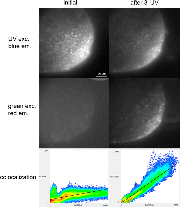
#DAPI and Hoechst dyes can be photoconverted with UV to forms that would be seen in green and red channels.
Avoid this issue by capturing channels in longer wavelengths first and/or by limiting UV exposure.
https://t.co/K97PdREHMH
@StillSciencing@FishCsCells Our patient data is anonymous once it reaches microscopy! Which is why this situation is a bit frustrating. They then to put everyone in the same bag 👎🏼
Dear microscopy tweeps,
We would like to purchase a core facility laptop for image analysis. Do any of you have any special spec recommendations? We mostly use Fiji, Qupath, Olympus software, MBF Bioscience software and Imaris...
#bioimaging#microscopy
@StillSciencing@FishCsCells Yup that’s it. I was once naive enough to go see IT about it 😆
The good news on that front is that we now have several platforms here that share our needs and finally have someone on the hospital administration looking into our special research IT needs. 🤷🏻♀️
@MixIndentations We tried for a while but we are considered a “commercial” account and we think investing in a laptop instead of a service like TeamViewer will be best
@FishCsCells We have a great workstation with all the goodies that we are not allowed to access from home because of the hospital network and no VPN 😭...
Today we've released our Imaging Facility Publication Guidelines!!!!
Designed to be displayed in #imaging#corefacilities, they aim to give recognition to our amazing #imagingscientist and acknowledgment to appropriate grants.
Please help us spread the word!
#UKLMF
Thank you everyone who showed to the virtual version of our annual image analysis workshops! It was great fun despite the technical issues 👍🏼
#Fiji#QuPath