Excited to share our new pre-print showcasing tools (chemical & optogenetic) for repositioning #vimentin in cells! Movie shows #ER (Halo-KDEL) sheets being relocalized together with vimentin (Vim) upon local vimentin pulling in U2OS. @gijsjekoenderi1
https://t.co/3VsESprJVn
Happy to share my cofirst-authorship in this work about my phD project👩🏻🔬.
We developed new reversible reporters to detect the dynamics of #MembraneContactSites and their associated #calciumsignaling.
Enjoy your read ⤵️ and our colourful pics & movies🎦.
https://t.co/B4Y7AIb18H
Pleased to present our latest work, where we show that the cell-cell adhesion protein afadin coopts mechanically-regulated elements of the cadherin-catenin complex to drive cytoskeletal engagement. https://t.co/SAmjUOuUHc
Which monomeric GFP is the brightest and most photostable?
A winner has just been announced, and it is mStayGold from the Miyawaki lab, edging out two other monomeric StayGold candidates.
https://t.co/x8cSa1B4Qu
We developed a new super-stable RFP for advanced microscopy, like CLEM, Rapid tissue clearing, Live 3D-SIM and live STED.
Read this #preprint on @researchsquare: mYongHong: a structure-guided design of stable and monomeric red fluorescent protein https://t.co/BUZYQAZhVC
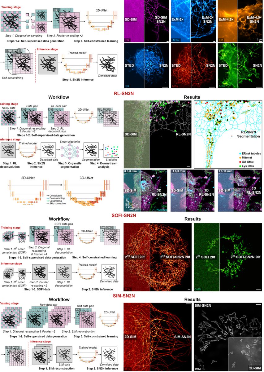
Out at @naturemethods just now!
https://t.co/SAiolzlnBy
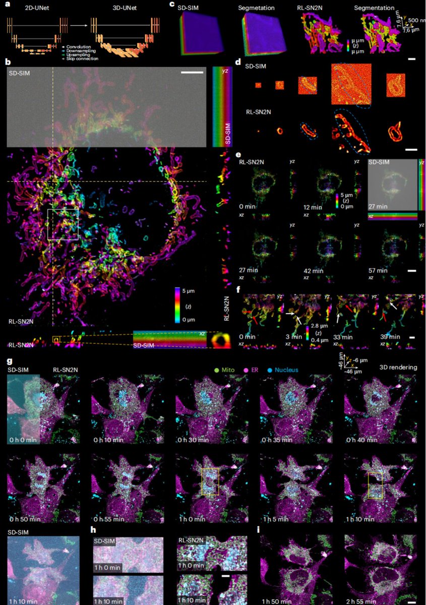
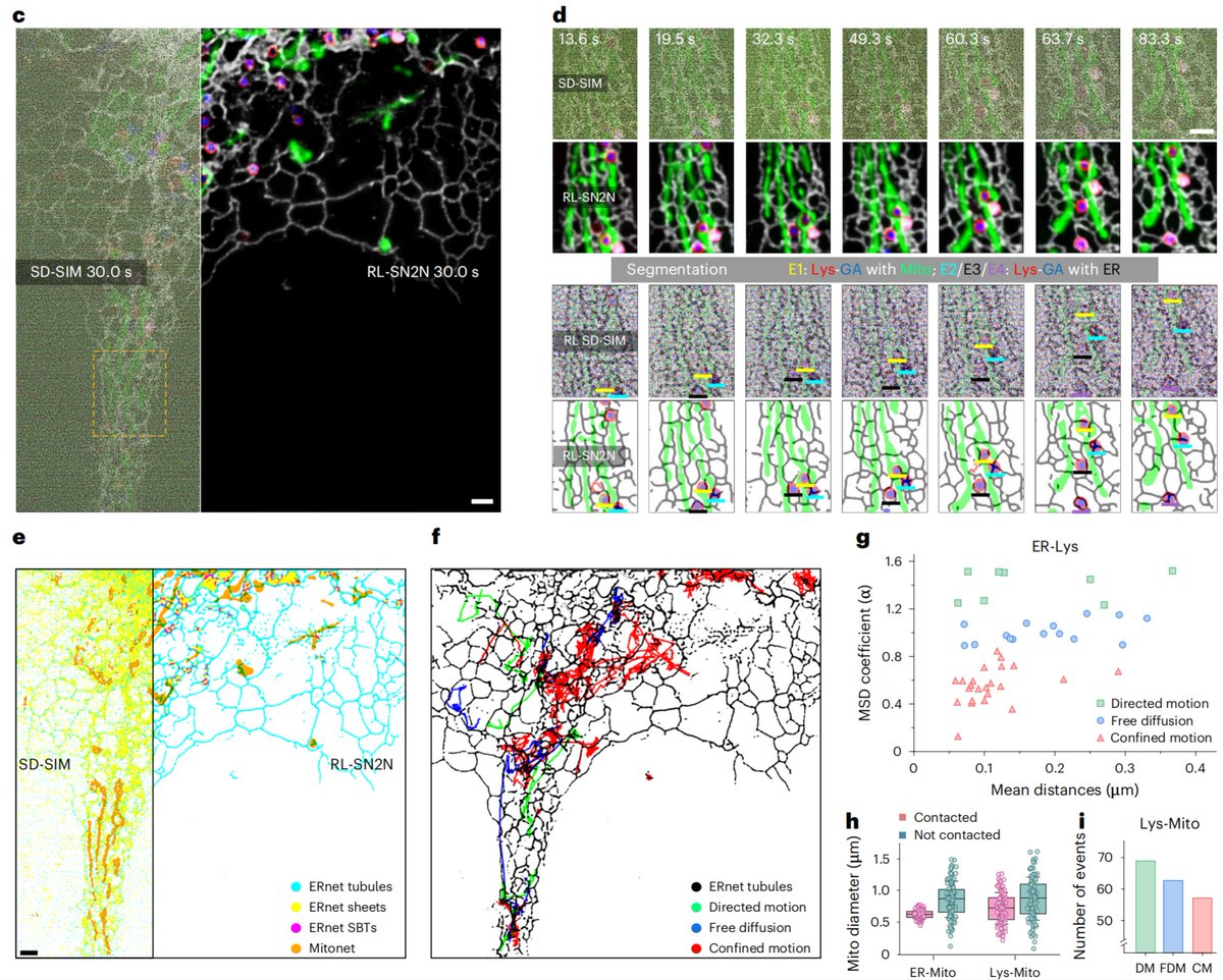
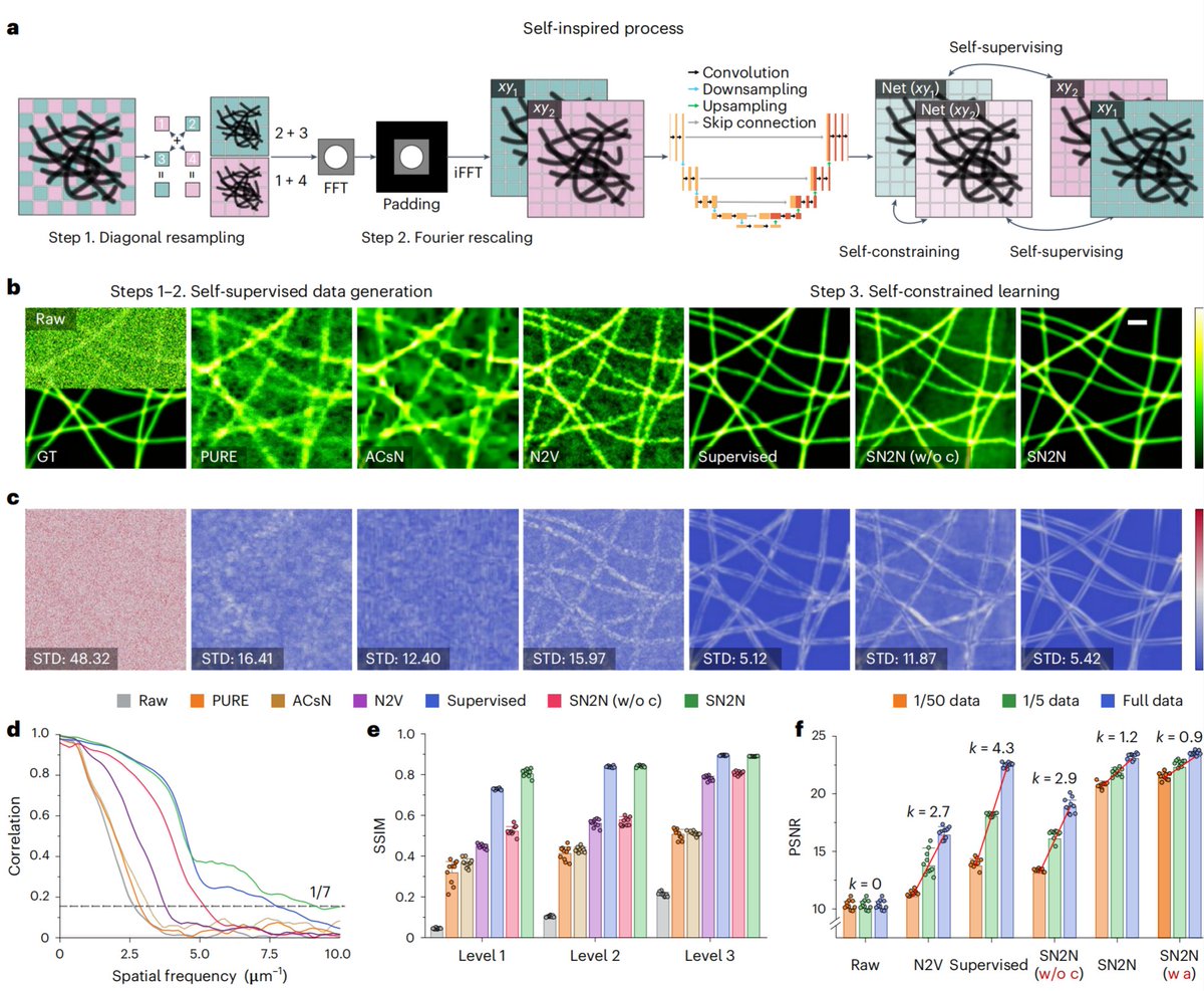
We present #SN2N, a flagship level unsupervised denoising solution. It’s fully competitive with supervised learning without needing large training-set and clean ground-truth, using 1 noisy frame for training.
(1/n)
Let me introduce: InstanSeg 🦠🔬💻👩🔬
This *would* have been a short thread about Thibaut Goldsborough’s PhD work… but he solved too many problems.
Now it's a long thread about 2 preprints, a whole new approach to cell segmentation & #opensource software to make it easy to use
BioRender updated their terms and, oh yes, I'm sure journals will be *thrilled* to print hyperlinks direct to BioRender's website at the bottom of figure legends.
https://t.co/fFHqxJRrou
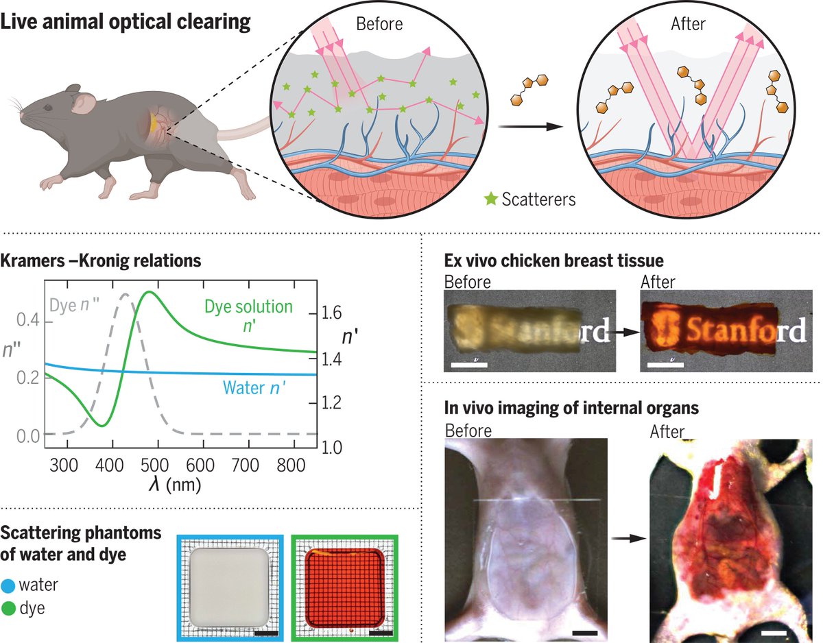
Long awaited (thanks @JamesDManton for posting the patent application!) and now out in @ScienceMagazine: Using absorption to look deeper by @HongNeuroTech - they achieved approx. 3 mm depth using the food dye tartrazine: #Microscopy
https://t.co/FkhBiQ1wDT
Scientific colour maps: yes!—But when and what not to do with them?
👉https://t.co/6shMBDbXLy
including resources for researchers, science communicators, software developers, publishing editors, and University faculty!
@ShepGracie@philipheron#DataVisualisation#UseBatlow
(1/x) We are excited to introduce VIPS (Volumetric Imaging of biological specimens via Photochemical Sectioning) in our preprint! VIPS uses a light-based process called “photochemical sectioning” to achieve petabyte-scale high/super-resolution imaging: https://t.co/Ubn7o4ocsa
@samjlord@sturm_gav The Visualization Toolset (from @kWolbachia) allows for adjusting the gamma of the current LUT through the use of the "Multi Tool" https://t.co/XKovhMaYxE
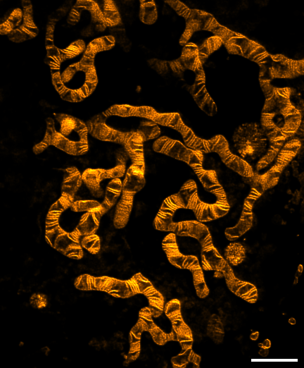
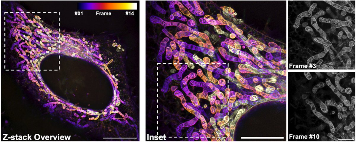
The new mitochondrial probe PKmito ORANGE FX in now available!
✅ Optimized for PFA or GA fixed cell mitochondria imaging
✅ Highly suited for STED imaging
✅ Compatible with immunostaining and CLEM
https://t.co/GPBcG2nt8m
Order it now from our distributors and take advantage of the 30% introductory discount.
Read more about PKmito ORANGE FX in the PNAS article by @ZhixingChen2 https://t.co/eIG6uFi0nW
Exciting preprint from the awesome folks (Mavrakis & Brasselet group) at @InstitutFresnel on new fluorescent reporters that will allow for polarisation measurements (in addition to regular microscopy) of actin dynamics in live cells!This is a big deal!
https://t.co/zBUaEgtDi7
After countless requests, we're thrilled to introduce PK Mito Orange FX - a fixable variant of PKMO. PKMO FX allows immunolabeling and CLEM approaches.
Happy to have contributed to this project led by @ZhixingChen2 and Christian Jüngst.
#mitochondria
https://t.co/nC6gX3LBGt