Congratulations to our #ACCFIT Jeopardy champions Team Dominican Republic! 🎉
Thank you for bringing your energy, expertise and competitive spirit to #ACCLATAM 2026. We’ll see you in Houston at #ACC27-#WCCardio!
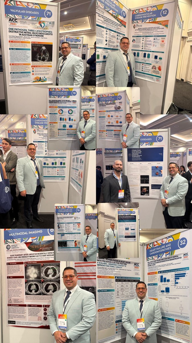
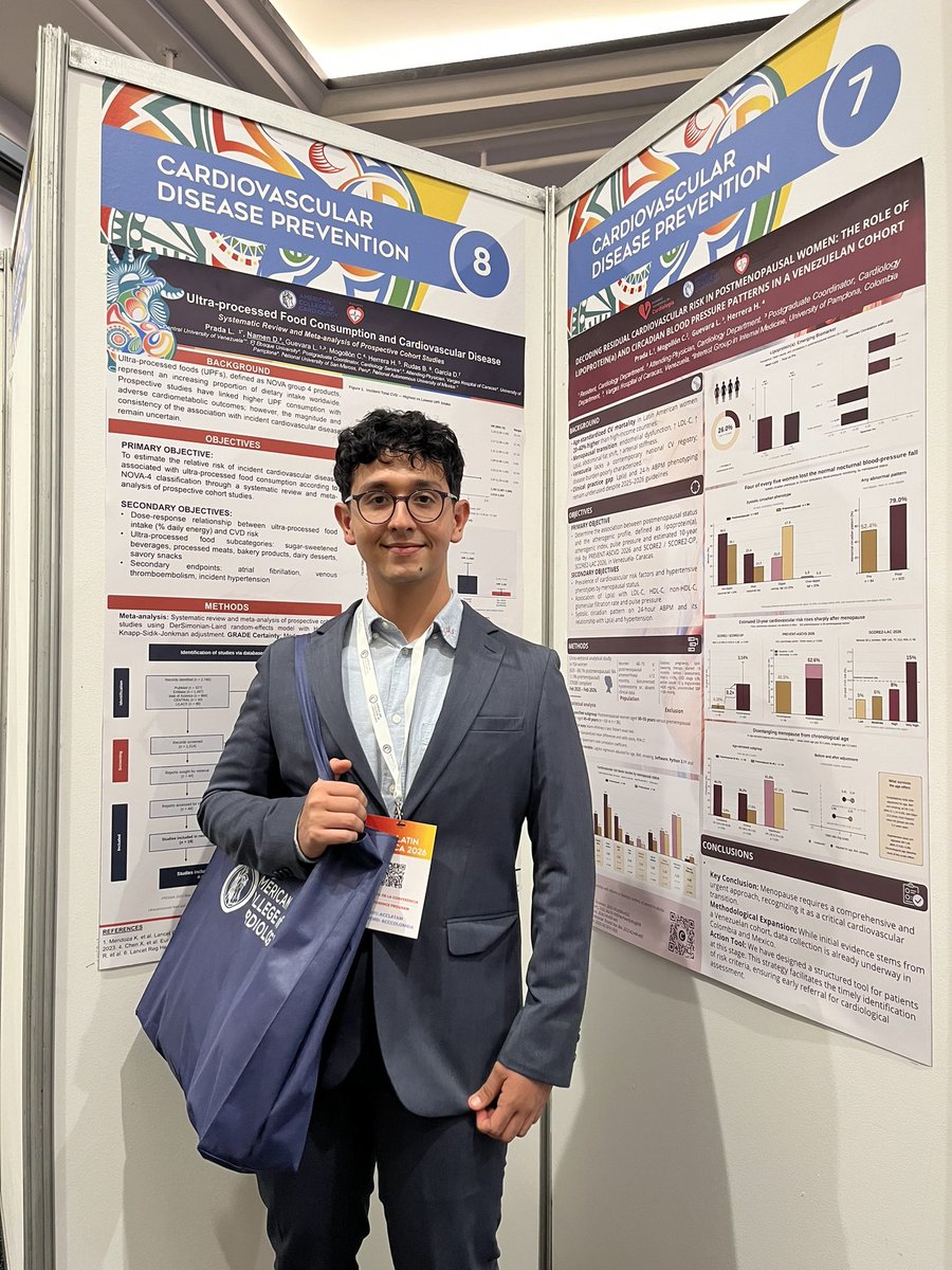
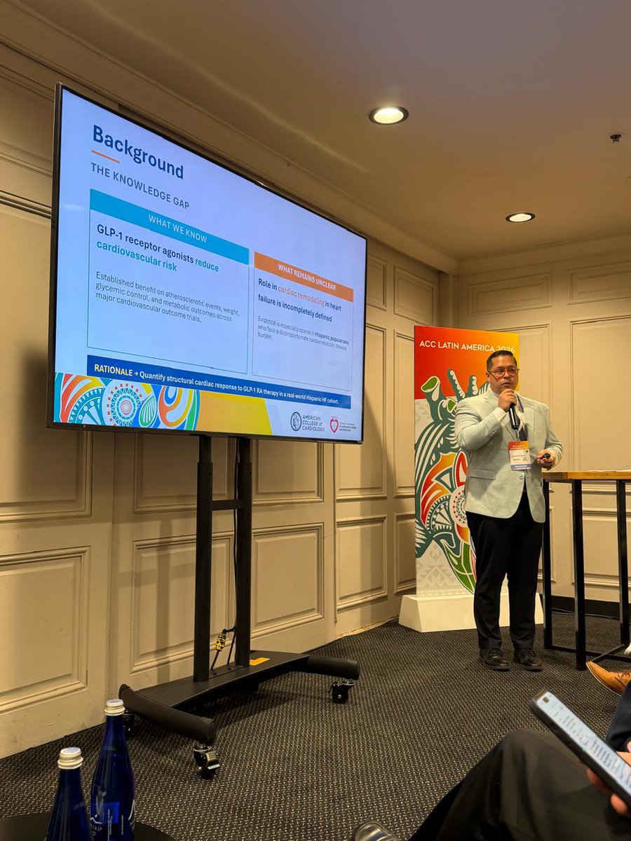
Late post for day 1 of #acclatam 2026 in Medellin. Get to know good people, and reconnect with great friends and colleagues. Proudly presented 10+ posters and 1 oral presentation of our significant study.
With a grateful heart, I am officially applying for Cardiology Fellowship. 🫀
This journey has been filled with sacrifice, uncertainty, growth, and the support of many people who believed in me along the way. I remain hopeful, humble, and deeply committed to becoming the cardiologist I have always dreamed of being.
AAMC ID: 13324466
#CardiologyFellowship #CardioTwitter #MedTwitter #Match2027 #IMG
#acclatinamerica2026
Grateful for the opportunity to learn from leading figures in cardiology. 🫀
Together with My Heart Team, proud to present three research projects and continue building the journey.
🔥 Hot off the press: We identified how and where AF begins.
I am excited to share this work.
Since the discovery of PV triggers more than 25 years ago, AF ablation has evolved into an empiric therapy centered on PVI. Yet, we have never been able to identify the arrhythmogenic substrate that determines why a premature beat initiates AF at one site and not another.
We believe this study changes that.
In this mechanistic study, we found that AF initiation arose predominantly through reentry originating from a small number of spatially discrete regions with steep repolarization gradients. Their distribution was unique to each patient, and they accounted for more than 92% of AF initiation episodes.
Importantly, the arrhythmogenic substrate was not defined by low voltage, and while conduction abnormalities were often present, they appeared to play a secondary role.
Its patient-specific distribution may help explain the controversy surrounding posterior wall isolation: the posterior wall harbored the substrate in only approximately 30% of patients. Importantly, in redo patients with durable PVI, right-atrial substrate (which is often beyond standard mapping strategies) was identified in 30%.
Most encouragingly, in an exploratory cohort of 24 patients with recurrent AF and durable PVI (50% PerAF), targeted elimination of these sites achieved 87.5% single-procedure freedom from any atrial tachyarrhythmia over a median of 489 days, without antiarrhythmic drugs.
The thoughtful accompanying editorial by Jon Kalman highlights the significance of this discovery and its central translational challenge of bringing it into real-world clinical practice.
But stay tuned! Technology is under development to transform this discovery into a rapid, automated functional mapping.
Current electroanatomic mapping relies largely on voltage amplitude and activation timing. We need to move beyond these measures and map how tissue actually functions - how it conducts, repolarizes, and responds to premature stimulation. This can add the critical dimension that has long been missing from substrate mapping and may reveal what truly makes cardiac tissue arrhythmogenic.
To me, this is the beauty of physiology: there are no black boxes. We can identify the sites that reproducibly initiate the arrhythmia, define their EP properties, eliminate them with ablation, and confirm that arrhythmia can no longer be initiated.
The next steps are prospective multicenter validation and technological development. Together, they may move AF ablation beyond empiric therapy toward truly individualized, mechanism-guided therapy.
📄 Paper in @JACCJournals:
https://t.co/izIR8HbNYk
📝 Editorial:
https://t.co/mYIilXkjJ7
#JACCCEP #AtrialFibrillation #CardiacElectrophysiology #CatheterAblation #EPeeps
Wide-complex tachycardia can be difficult to classify, but the limb lead algorithm provides a quick, stepwise approach using only the limb leads.
How it works:
Step 1: Is there a monophasic R wave in lead aVR?
If yes, the rhythm is ventricular tachycardia (VT).
Step 2: If not, are leads I, II, and III all predominantly negative?
If yes, this strongly favors VT.
Step 3: If neither criterion is present, examine the inferior leads (II, III, aVF). If they are all monophasic positive or all monophasic negative, check the remaining limb leads (I, aVR, aVL) for opposite polarity. If this pattern is present, the diagnosis is VT.
🟥 If these criteria are not met, the algorithm does not classify the rhythm. Additional ECG criteria and clinical assessment are required to distinguish VT from supraventricular tachycardia (SVT) with aberrant conduction.
Take-home message:
The limb lead algorithm is a practical bedside tool that can rapidly identify many cases of VT using only limb leads. However, it is not designed to diagnose every wide-complex tachycardia, so an indeterminate result should prompt a comprehensive 12-lead ECG analysis rather than exclusion of VT.
Reference: Zipes DP, Jalife J. Cardiac Electrophysiology: From Cell to Bedside.