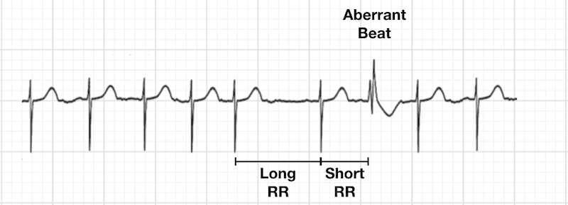
✅هذا هو الـ Ashman's phenomenon، و هي ظاهرة كهربائية تصير في القلب وتلاحظها وانت تقرا الـ ECG، وأساسها يرجع لمفهوم اسمه aberrant conduction، يعني توصيل كهربائي غير طبيعي داخل الـ ventricles بس مو بسبب مشكلة عضوية في القلب، إنما بسبب اختلاف في الـ refractory period بين الـ bundle branches، ف عشان تفهمها لازم تعرف إن الـ right bundle branch عنده refractory period أطول من الـ left bundle branch بشكل طبيعي، يعني يحتاج وقت أطول عشان يرجع "يتعافى" ويصير جاهز يوصل نبضة كهربائية جديدة، ف لما يصير عندك نبضة قصيرة (short cycle) تجي بعد نبضة طويلة (long cycle)، يعني فيه اختلاف واضح بين طول الـ R-R intervals المتتالية، هذا التغيير المفاجئ يخلي الـ right bundle branch لسا في فترة الـ refractory period بينما الـ left bundle branch خلاص تعافى وجاهز، ف النبضة الجديدة تنتقل عن طريق الـ left bundle branch بس وتوصل متأخرة للـ right side.
هذا يعطيك شكل على الـ ECG يشبه تماما الـ right bundle branch block (RBBB) morphology يعني QRS عريض مع شكل معين في الـ V1 يشبه rSR' وتغيرات بالـ T wave، وأهم شي تعرفه إن هذا الشكل مؤقت وما يمثل مشكلة حقيقية أو دائمة في نظام التوصيل.
📌لو حاب تفهم الحالة اكثر، الفيديو هذا يشرحها بطريقة جميلة: https://t.co/7QQ4mJOR7W
Two young patients with congenital mitral valve disease. Two A4C. Two PLAX.
Mitral arcade 🆚 parachute MV
Match each A4C to its corresponding PLAX.
Our 👀 see what our 🧠 already knows #echofirst
M-Mode Echo: A Window into LV Function
This schematic illustrates how 2D-guided M-mode echocardiography measures left ventricular (LV) wall thickness and chamber dimensions throughout the cardiac cycle.
Timing of Measurements:
- Diastole: Measured at the Q wave of the ECG
- Systole: At max posterior septal motion, when motion is normal
Key Parameters:
- LVIDd = LV internal diameter in diastole
- LVIDs = LV internal diameter in systole
- STd/STs = Septal thickness (diastole/systole)
- PWTd/PWTs = Posterior wall thickness (diastole/systole)
What is Fractional Shortening (FS)?
Fractional shortening (FS) is the percentage change in LV internal diameter from diastole to systole.
Formula
FS (%) = [(LVIDd − LVIDs) ÷ LVIDd] × 100
Normal range: 25–45%
Clinical Significance:
- Reduced FS% suggests impaired LV systolic function (e.g., in heart failure or cardiomyopathy)
- Increased wall thickness may point to LV hypertrophy (e.g., due to hypertension or aortic stenosis)
- Rapid and reproducible, ideal for screening and serial assessments
Source: Aurigemma GP et al., Curr Probl Cardiol. 1995;20:381.






![TrackYourHeart's tweet photo. M-Mode Echo: A Window into LV Function
This schematic illustrates how 2D-guided M-mode echocardiography measures left ventricular (LV) wall thickness and chamber dimensions throughout the cardiac cycle.
Timing of Measurements:
- Diastole: Measured at the Q wave of the ECG
- Systole: At max posterior septal motion, when motion is normal
Key Parameters:
- LVIDd = LV internal diameter in diastole
- LVIDs = LV internal diameter in systole
- STd/STs = Septal thickness (diastole/systole)
- PWTd/PWTs = Posterior wall thickness (diastole/systole)
What is Fractional Shortening (FS)?
Fractional shortening (FS) is the percentage change in LV internal diameter from diastole to systole.
Formula
FS (%) = [(LVIDd − LVIDs) ÷ LVIDd] × 100
Normal range: 25–45%
Clinical Significance:
- Reduced FS% suggests impaired LV systolic function (e.g., in heart failure or cardiomyopathy)
- Increased wall thickness may point to LV hypertrophy (e.g., due to hypertension or aortic stenosis)
- Rapid and reproducible, ideal for screening and serial assessments
Source: Aurigemma GP et al., Curr Probl Cardiol. 1995;20:381.](https://pbs.twimg.com/media/HKSfSmnbAAAI-Sq.jpg)