This video shows the application of a Mayfield-type skull clamp to rigidly immobilize the patient’s head before neurosurgery. The three-pin fixation device locks the skull in place, preventing even millimeter-scale movement that could compromise precision during brain procedures. The MRI at the end displays the intracranial lesion (likely a tumor or similar pathology) that the surgery will address. Head fixation is a standard, critical step in most intracranial operations.
Suboccipital muscles are a group of four small muscles located just under your skull. They consist of two rectus and two obliquus muscles.
Together, these muscles help control fine movements and stabilization of the head.
What happens in the neck in cervical myelopathy?
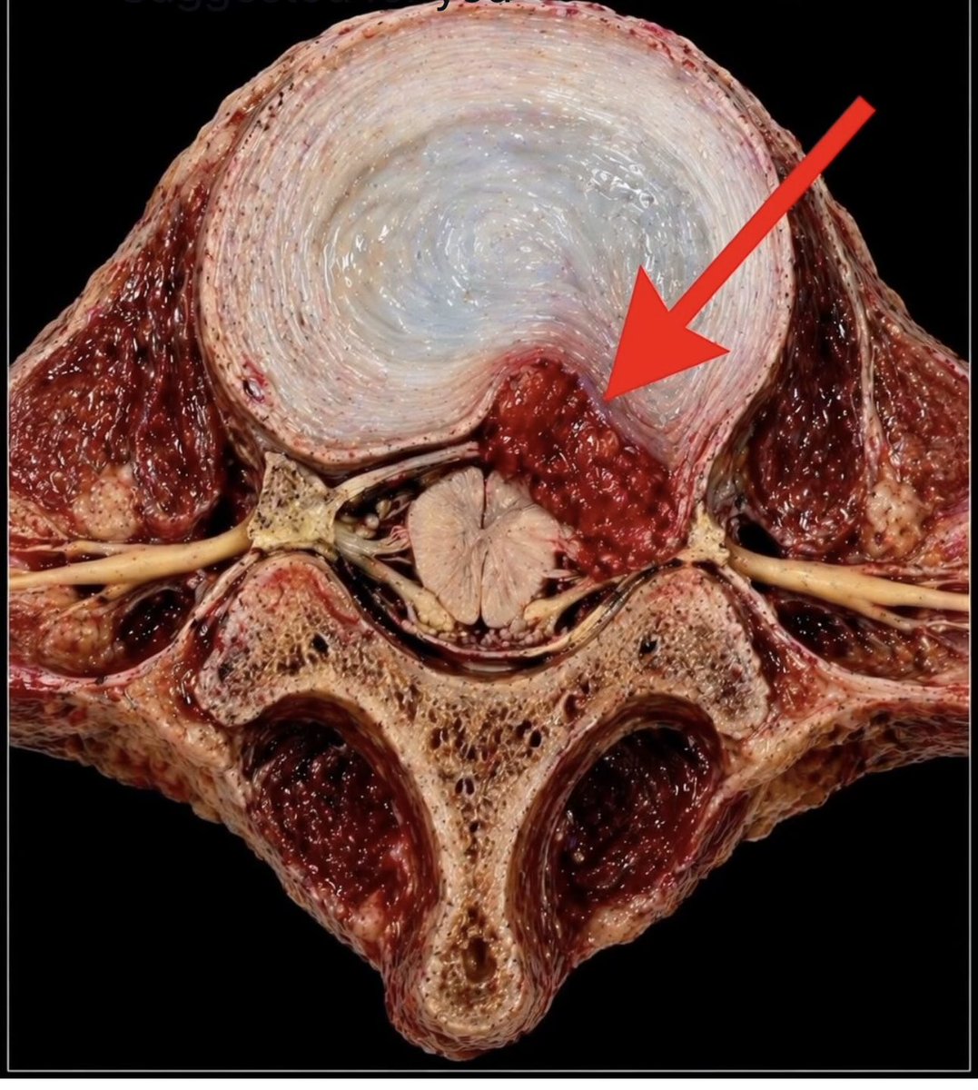
As we get older, the spine can slowly change. The discs lose height, bones can form extra edges, and ligaments may thicken or even turn into bone. Over time, these changes can make the space around the spinal cord smaller.
When that space narrows, the spinal cord can become compressed. This may lead to symptoms like pain, weakness, numbness, or trouble with balance and walking.
These changes are often described as “degeneration” on a scan. But for patients, the effects can be very real and can worsen if not recognized early.