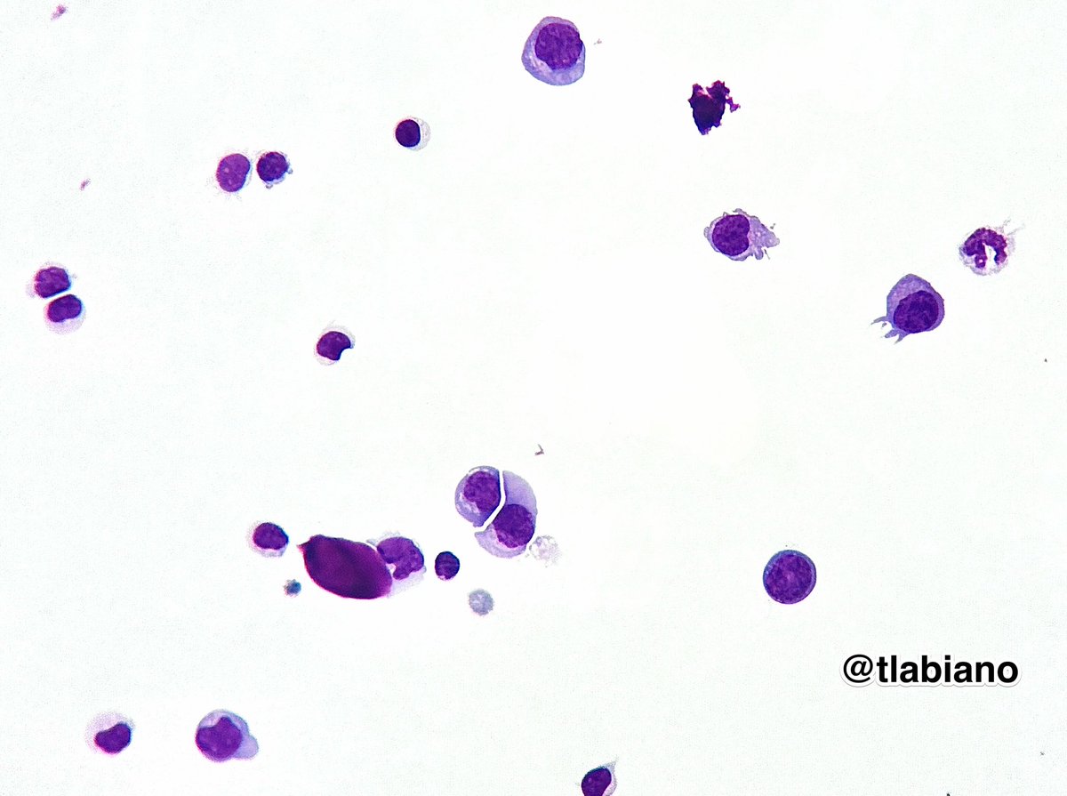
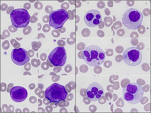
🧠💧Cerebrospinal fluid #CSF#cytology 🔬Large cells w/ pleomorphic population of lymphoid cells with irregular nuclear contours.
📋✍🏼Patient has history of Diffuse large B-cell lymphoma.
#giemsa#cytopath
Something’s brewing in the bone marrow…
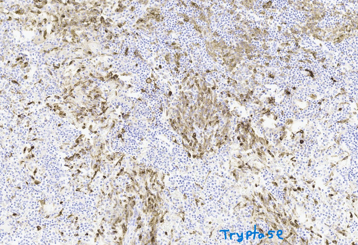
Don’t Tryp on these mast cells!
A spindling review of systemic mastocytosis:
🚨Atypical= Spindled
🔴Cluster > 15 cells
🦴Para trabecular/perivascular
🧪CD2+ CD25+ Tryptase+
🧬KIT D816V
#hemepath#PathX#ABPath
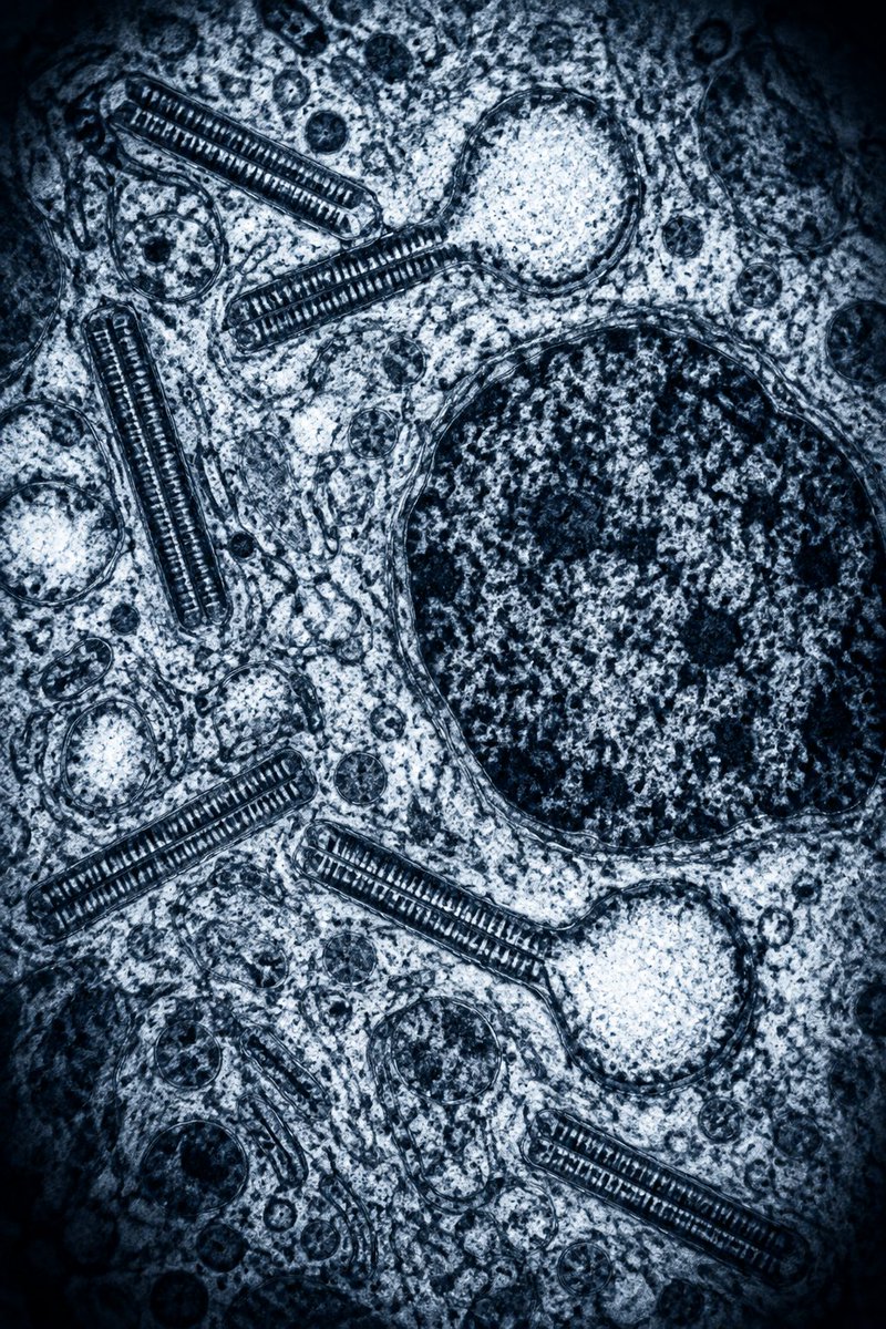
A characteristic “tennis-racket” cytoplasmic organelle is identified on electron microscopy.
In which condition is this finding classically seen?
A. Malignant histiocytosis
B. Langerhans cell histiocytosis
C. Juvenile xanthogranuloma
D. Niemann–Pick disease
The CHIP/CCUS field is becoming the new MGUS. Stop ordering NGS on all mild cytopenias. Follow them for a few months. If it’s worse than do what you want.
🩸 Red-Cell Antigen Phenotyping: How to Interpret It
🔬 Antigen phenotyping determines which inherited antigens are present (+) or absent (−) on the patient’s red blood cells.
🧬 Major systems commonly tested:
🔴 Rh: C, c, E, e
🟠 Kell: K, k
🟡 Duffy: Fya, Fyb
🟢 Kidd: Jka, Jkb
🔵 MNS: M, N, S, s
✅ Positive antigen = expressed on the RBCs
❌ Negative antigen = absent; the patient may form an alloantibody against it after exposure through transfusion or pregnancy.
⚠️ Examples:
E− → risk of developing anti-E
K− → risk of developing anti-K
Fya− → risk of developing anti-Fya
Jka− → risk of developing anti-Jka
🩺 Why is phenotyping important?
🩸 Investigating or preventing RBC alloimmunization
🎯 Selecting antigen-negative compatible units
♻️ Supporting chronically transfused patients
🔎 Interpreting complex antibody panels
🛡️ Preventing delayed hemolytic transfusion reactions
🤰 Assessing antibodies relevant to hemolytic disease of the fetus and newborn
🌍 Special example: Fy(a−b−)
This is the Duffy-null phenotype, common among individuals of African and Middle Eastern ancestry.
🧪 It may be associated with a persistently lower circulating absolute neutrophil count—now called Duffy-null–associated neutrophil count (DANC).
✅ Usually no increased infection risk
✅ Does not require treatment by itself
⚠️ Diagnose only after clinical correlation and exclusion of other relevant causes of neutropenia.
🚨 Important limitations
🩸 Phenotyping may be unreliable following recent transfusion because circulating donor RBCs can produce mixed results.
🧪 A positive direct antiglobulin test may also interfere with serologic testing.
🧬 RBC genotyping is preferable when recently transfused, DAT-positive, extensively alloimmunized, or when serologic findings are inconclusive.
📌 Phenotype tells us what is expressed on circulating RBCs; genotype predicts inherited antigen expression.
🔗 References:
American Society of Hematology—Duffy-null associated neutrophil count
NCBI—Blood Groups and Red Cell Antigens
Canadian Blood Services—Serological best practices
#Hematology #TransfusionMedicine #BloodBank #DuffyNull #DANC
Anybody, any experience with Adzynma for congenital TTP? Any pros and cons. Personal experience?
For those who are not aware about Adzynma,
Adzynma (apadamtase alfa) is a recombinant enzyme replacement therapy used to treat both children and adults with congenital thrombotic thrombocytopenic purpura (cTTP), a rare inherited blood-clotting disorder. It replaces the missing ADAMTS13 enzyme to prevent harmful blood clots, low platelets, and anemia.
#cTTp #adzynma
Ps. There is no conflict of interest.
🧵 Rosai–Dorfman–Destombes disease (RDD): hematology pearls
1️⃣ What is it?
A rare histiocytic neoplasm formerly called sinus histiocytosis with massive lymphadenopathy.
📌 Derived from activated macrophage-lineage histiocytes
📌 Usually indolent, but behavior ranges from spontaneous remission to organ-threatening disease.
2️⃣ Classic presentation
👤 Children or young adults—although any age is possible
🦒 Massive, painless, bilateral cervical lymphadenopathy
🌡️ Fever, night sweats or weight loss
🩸 Neutrophilia, anemia, raised ESR/CRP
🧪 Polyclonal hypergammaglobulinemia
3️⃣ Extranodal disease
Occurs in approximately 40% of patients:
🩹 Skin—most frequent extranodal site
👃 Nasal cavity/sinuses
👁️ Orbit
🦴 Bone
🧠 Dural/CNS masses mimicking meningioma
🫁 Airways/lung
🫘 Kidney
🧬 Salivary gland, breast, testes and soft tissue
⚠️ Airway, CNS, orbital, renal or multiorgan disease may become organ-threatening.
4️⃣ Pathology hallmark
🔬 Large pale histiocytes showing emperipolesis: intact lymphocytes, plasma cells or neutrophils within the histiocyte cytoplasm without destruction.
Immunophenotype:
✅ S100 positive
✅ CD68/CD163 positive
✅ OCT2 and cyclin D1 frequently positive
❌ CD1a negative
❌ Langerin/CD207 negative
📌 Emperipolesis is supportive but not entirely specific—clinicopathological correlation is essential.
5️⃣ Molecular biology
🧬 Somatic MAPK-pathway alterations occur in a subset:
• KRAS
• NRAS
• MAP2K1
• ARAF
• CSF1R
• Rare BRAF alterations
➡️ Perform tissue next-generation sequencing in severe, refractory or disseminated disease because it may identify a target for MEK inhibition.
6️⃣ Clinical categories
🔵 Classical nodal RDD
🟣 Extranodal RDD
🧬 Familial RDD—consider SLC29A3-related disease
🛡️ Immune-associated RDD: SLE, autoimmune cytopenias or ALPS
🎗️ Neoplasia-associated RDD: may coexist with lymphoma, myeloid neoplasms or other cancers
7️⃣ Differential diagnosis
🔍 Langerhans-cell histiocytosis
🔍 Erdheim–Chester disease
🔍 Juvenile xanthogranuloma
🔍 Hodgkin/non-Hodgkin lymphoma
🔍 IgG4-related disease
🔍 Sarcoidosis
🔍 Tuberculosis or fungal infection
🔍 Metastatic carcinoma/melanoma
🔍 Hemophagocytic lymphohistiocytosis
⚠️ Increased IgG4-positive plasma cells alone do not establish IgG4-related disease.
8️⃣ Initial work-up
🧫 Core or excisional biopsy with expert hematopathology review
🩸 CBC, renal/liver profile, ESR/CRP and immunoglobulins
🛡️ Autoimmune testing when clinically indicated
📸 FDG-PET/CT or CT neck/chest/abdomen/pelvis
🧠 MRI for neurological, orbital or sinonasal disease
🧬 NGS for MAPK-pathway alterations
🦴 Bone marrow only for unexplained cytopenias or suspected marrow involvement
9️⃣ Who can be observed?
👀 Observation is appropriate for:
✅ Asymptomatic nodal or cutaneous disease
✅ Non-bulky disease without organ compromise
✅ Stable postoperative residual disease
Many cases remain stable or regress spontaneously. Reassess clinically and radiologically according to the involved sites.
🔟 When to treat?
🚨 Progressive or symptomatic disease
🚨 Vital-organ dysfunction
🚨 Airway obstruction
🚨 Neurological or visual compromise
🚨 Renal involvement
🚨 Severe autoimmune cytopenia
🚨 Multifocal/disseminated disease affecting quality of life
1️⃣1️⃣ Treatment approach
🔪 Surgery: preferred for isolated, accessible or compressive lesions; complete excision may be curative.
💊 Corticosteroids: prednisone approximately 40–70 mg/day followed by an individualized taper; responses may be transient.
☢️ Radiotherapy: selected unresectable, residual or critical-site disease.
🧪 Cladribine: 5 mg/m²/day IV for 5 days every 28 days, commonly for 3–6 cycles in severe or refractory systemic disease; monitor for prolonged myelosuppression and opportunistic infection.
Other selected options include low-dose methotrexate ± 6-mercaptopurine, sirolimus—particularly with immune dysregulation—and lenalidomide-based therapy.