Team care. Grateful to be part of the sports medicine for the Texans NFL at the NFL Combine 2025 #Texans, #NFLcombine2025#mskradiology
Thankful for the opportunity to be part of the scientific program of the annual #NFLPS
Exciting week in Indy!
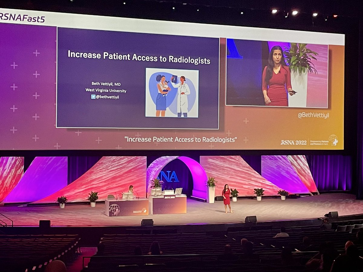
My talk #RSNAFast5 - inspired by 2 giants in Radiology.
Collaborative Radiology Clinic model: Dr. Nicks @MKMSKER at @UTHradiology
Dedicated Radiology Clinic model: Dr. Avneesh Chhabra @AChhabraMD at @UTSW_Radiology
Increased patient access to radiologists? It’s already on!!
Don’t get left out in the cold! 🥶
Beginning Jan 1, 2022 the RSNA Case Collection will be available only to members or by subscription. Subscribe today to use this valuable resource and point-of-care tool!
🔗https://t.co/5UrS1o1oHU
@RSNA#RSNACC#RadEd
🎄The answer is Osteitis condensans Ilii, Fir sure!
https://t.co/TmIjXFUaSa
Thanks #RSNACC#MSKRad Dep Ed Dr Kumaravel @MKMSKER@UTRadRes & authors Drs Paulo Miro & Jimmy Saade!
@DMG_AZ@uazmedphx
🔔 Happy Holidays and warm wishes for the New Year from @RCC_Editor@RSNA 🔔
How NOT to miss a compression fracture of the vertebral body on CT?
Look for the following features: 5Ds
1. Dense (sclerotic) # line
2. Depression- wedging
3. Deformity- end plates (depression)
4. Deformity- buckling or cortical step
5. Discontinuity- cortex (corners)
Another excellent Emergency session by @MKMSKER on imaging of soft tissue injuries of ankle and how subtle # and soft tissue edema can reveal a significant injury. Avulsion # represents tip of the iceberg.
@ASER_ERad
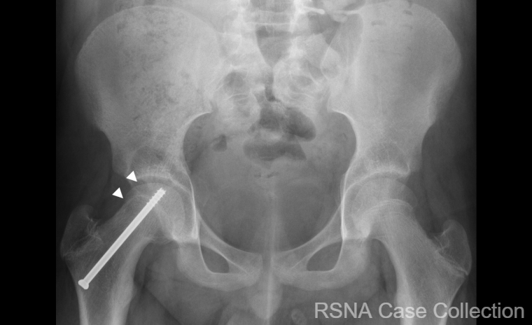
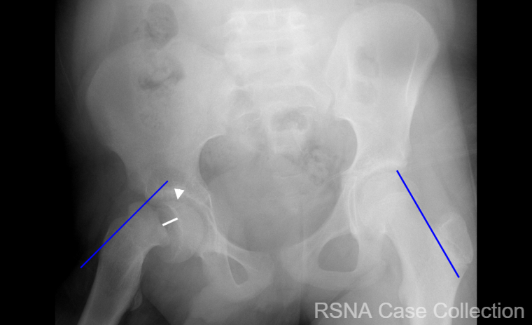
Avulsion of the gluteus maximus tendon insertion is an unusual associated injury to find during acetabular fracture ORIF - it’s easily diagnosed and then repaired via the KL exposure - suture anchors were used for this one.
🎈Here's to celebrating a successful first year of the RSNA Case Collection #RSNACC! Our collection now has over 600 high quality cases. Sincere thanks to our amazing editors, authors and staff! We look forward to another great year🎈
@RCC_Editor@moshirimd@RSNA
A true honor to be inducted as a fellow of the @RadiologyACR#FACR#ACR2021 . Unfortunately, it was all virtual but I was thrilled to share the moment with my mom in India over YouTube. Thanks to all my mentors and @TxRadSociety for the support
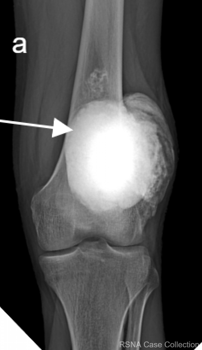
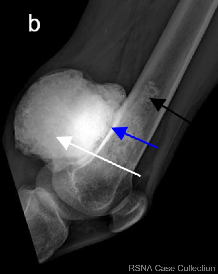
Make no “bones” about it- the diagnosis for this #RSNACC COW is Parosteal Osteosarcoma!! https://t.co/MgiJxpb9im
Thanks @RSNA#RSNACC#MSKRad Deputy Ed Dr Kumaravel @MKMSKER@UTRadRes & authors Drs Richard-Edwards, Sood and Ncube @VisheshSood88@ivncube83 Groote Schuur Hospital
Honored to be part of the #AUR2021 Management course as faculty with @NBeckmann_MD . Thanks for letting us present our case. Learned a lot from current and future leaders. @AURtweet
Palmer class 1B lesion with complete detachment of the ulnar insertion of the TFCC, and secondary distal radioulnar joint instability. Diagram and arthroscopic image show the reattachment of the TFCC to the foveal insertion. Follo-up MR image demonstrates complete TFCC healing
Although disappointing for not being in person for the #NFLcombine2021 in Indy, still managed to review all imaging of prospective #NFLDraft players. Remote radiology at work!
Exciting to see #NFLDraft2021 in progress.
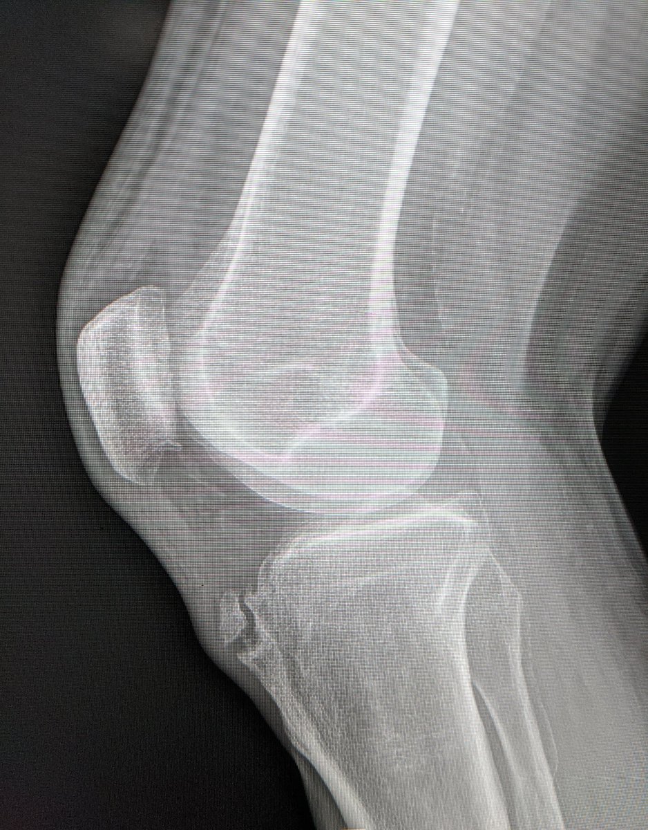
43 year old presenting with anterior knee pain.
Tibial tuberosity edema.
Edema across the fibrous union synchondrosis to the native tibia. Fluid in pretibial bursa.
Diagnosis: Adult presentation of Osgood Schlatter disease.
#FOAMed#MSKrad#sportsmedicine
Acetabular dysplasia commonly produces acetabular chondral flaps. Abnormal shear forces due to anterolateral femoral head subluxation results in a central tear in the articular cartilage and a contiguous chondrolabral sleeve that extends peripherally (‘‘ inside-out ’’ mechanism)